
Image
|
Figure Caption
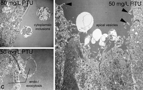
Fig. 5
Apical alterations in zebrafish thyrocytes caused by propylthiouracil exposure. At concentrations ≥10 mg/L, cytoplasmic inclusions were evident (A). At concentrations ≥2.5 mg/L, numerous apical vesicles were seen protruding into the follicular lumen (B). Bleb-like structures indicate endo- or exocytotic processes at concentrations ≥10 mg/L (B, C). Moderate proliferation of microvilli can be observed at concentrations ≥10 mg/L (▸; B). Magnifications: A: 12,500×; B: 8,000×; C: 20,000×.
Acknowledgments
This image is the copyrighted work of the attributed author or publisher, and
ZFIN has permission only to display this image to its users.
Additional permissions should be obtained from the applicable author or publisher of the image.
Full text @ Toxicol. Pathol.