
- Title
-
Glucocorticoid receptor, but not mineralocorticoid receptor, mediates cortisol regulation of epidermal ionocyte development and ion transport in zebrafish (danio rerio)
- Authors
- Cruz, S.A., Lin, C.H., Chao, P.L., and Hwang, P.P.
- Source
- Full text @ PLoS One
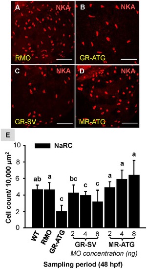
Effect of corticosteroid receptor gene knockdown on NaRC number. Zebrafish embryos at the 1~4 cell-stage were microinjected with glucocorticoid receptor ATG-MO (GR-ATG), GR-splice variant MO (GR-SV), mineralocorticoid receptor ATG-MO (MR-ATG), or Random-MO (RMO; control). Representative images of yolk-sac NaRCs labeled with anti-α sub-unit of Na+-K+-ATPase (NKA) in RMO (A), GR-ATG (B), GR-SV (C), and MR-ATG (D) morphants. NaRC numbers are compared in (E). Values are presented as the mean ±s.d. (n = 10–12). abIndicates statistically significant differences (<0.05) between groups as determined by one-way ANOVA (Tukey’s pair-wise comparison). Scale bar: 100 μm (A–I). |
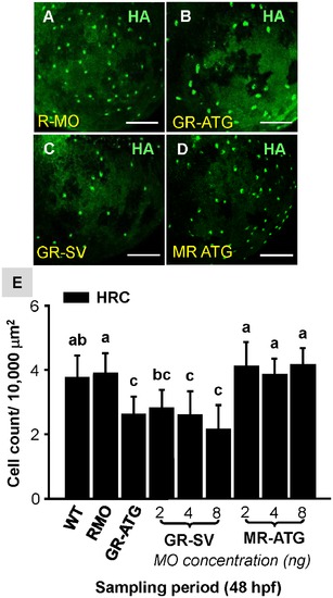
Effect of corticosteroid receptor gene knockdown on HRC number. Zebrafish embryos at the 1~4 cell-stage were microinjected with glucocorticoid receptor ATG-MO (GR-ATG), GR-splice variant MO (GR-SV), mineralocorticoid receptor ATG-MO (MR-ATG), or Random-MO (RMO; control). Representative images of yolk-sac HRCs labeled with H+-ATPase (HA) in RMO (A), GR-ATG (B), GR-SV (C), and MR-ATG (D) morphants. HRC numbers are compared in (E). Values are presented as the mean ±s.d. (n = 10–12). abIndicates statistically significant differences (<0.05) between groups as determined by one-way ANOVA (Tukey’s pair-wise comparison). Scale bar: 100 μm (A–F). |
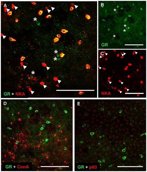
Localization patterns of GR protein in epidermal cells of zebrafish embryos. Representative images of the yolk-sac of wild-type embryos (48–96 hpf) labeled with anti-glucocorticoid receptor (GR) and anti-α sub-unit of N+-K+-ATPase (NKA) (a marker for NaRCs) (A), anti-GR (B), anti-NKA (C), anti-GR and ConA (a marker for HRCs) (D) and anti-GR with anti-p63 (a marker for epidermal stem cells) (E). Arrow heads indicate NaRCs without a GR signal; asterisks indicate GR-expressing cells without an NKA signal (A–C). Scale bar: 100 μm (A–E). |
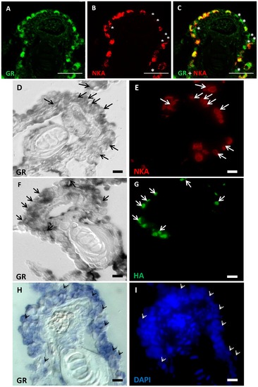
Expression patterns of GR mRNA and protein in the ionocytes of adult zebrafish gills. Representative images of gill paraffin sections labeled with anti-glucocorticoid receptor (GR) (A), anti-alpha sub-unit of N+-K+-ATPase (NKA) (a marker for NaRCs) (B), and anti-GR and anti-NKA (C). Asterisks indicate gr-expressing cells without an NKA signal (B–C). Gill cryosections reveal co-localization of gr mRNA with NKA (D–E) and anti-H+-ATPase (HA) (F–G). After gr mRNA in situ hybridization, nuclear/cellular structure can be visualized with DAPI signals (H–J). Arrows and arrow-head indicate cells with colocalized signals (D–G). Scale bars: 25 μm (A–C) and 5 μm (D–I). EXPRESSION / LABELING:
|

Unillustrated author statements PHENOTYPE:
|