
Image
|
Figure Caption
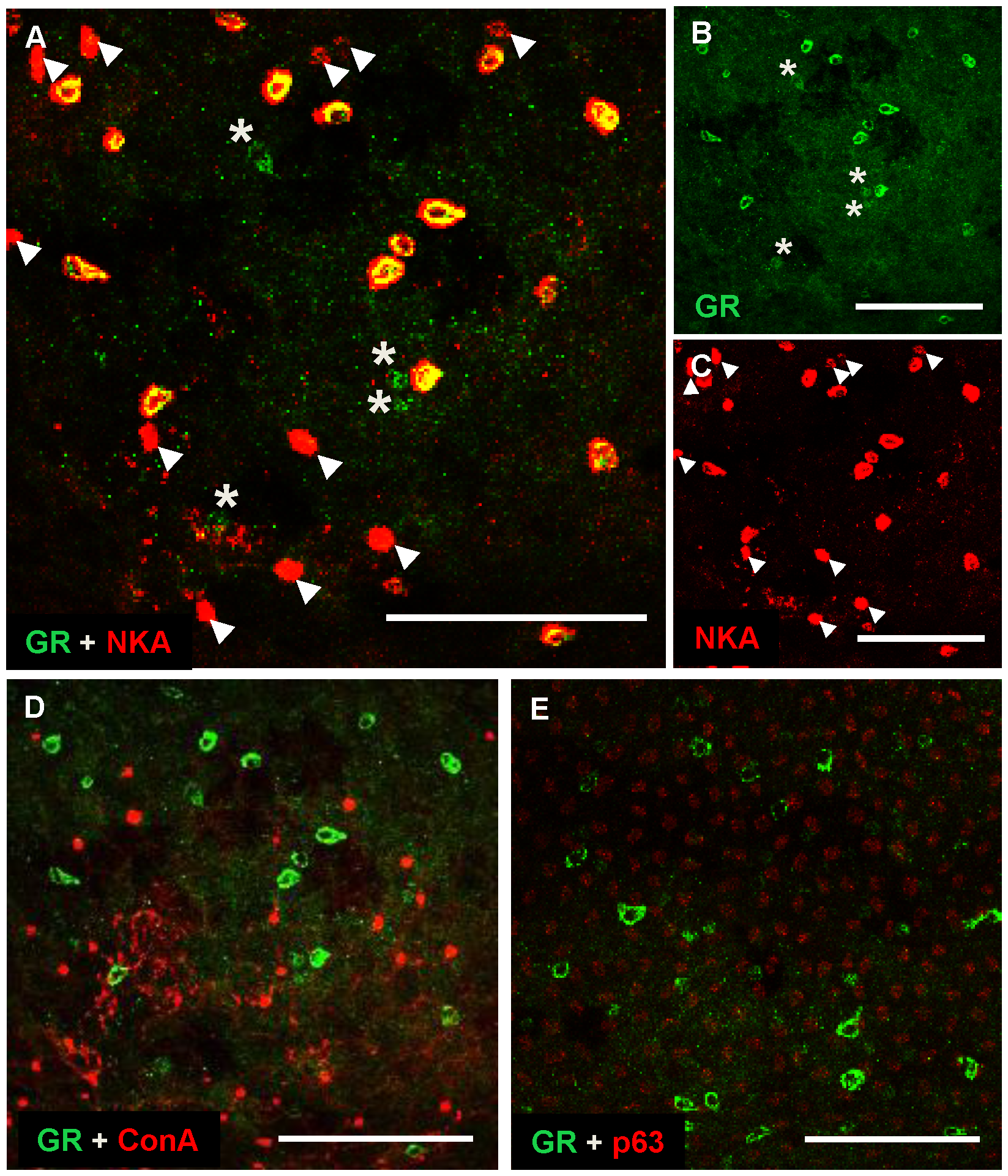
Fig. 7
Localization patterns of GR protein in epidermal cells of zebrafish embryos.
Representative images of the yolk-sac of wild-type embryos (48–96 hpf) labeled with anti-glucocorticoid receptor (GR) and anti-α sub-unit of N+-K+-ATPase (NKA) (a marker for NaRCs) (A), anti-GR (B), anti-NKA (C), anti-GR and ConA (a marker for HRCs) (D) and anti-GR with anti-p63 (a marker for epidermal stem cells) (E). Arrow heads indicate NaRCs without a GR signal; asterisks indicate GR-expressing cells without an NKA signal (A–C). Scale bar: 100 μm (A–E).
Figure Data
Acknowledgments
This image is the copyrighted work of the attributed author or publisher, and
ZFIN has permission only to display this image to its users.
Additional permissions should be obtained from the applicable author or publisher of the image.
Full text @ PLoS One