
|
Fig. 8
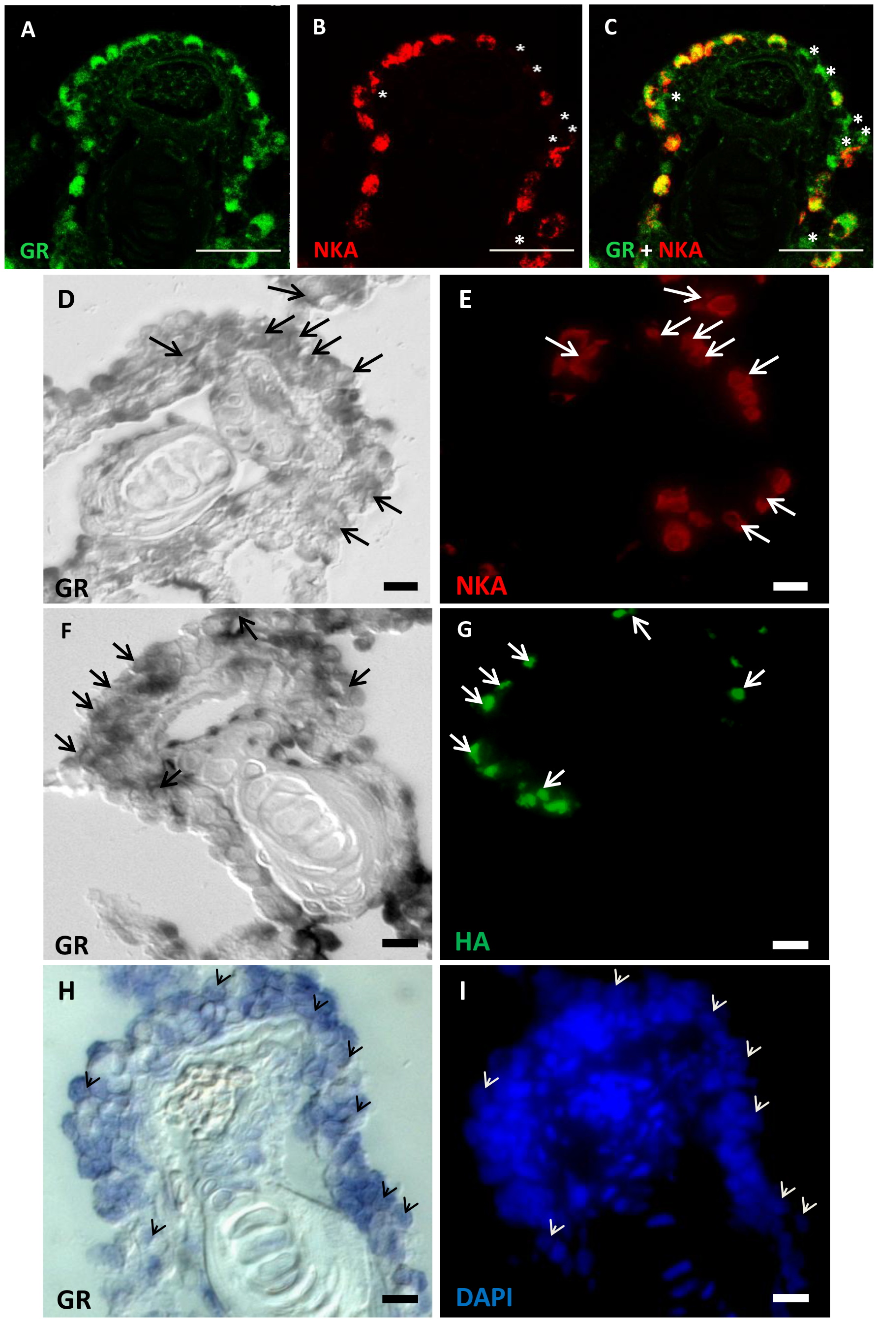
Expression patterns of GR mRNA and protein in the ionocytes of adult zebrafish gills.
Representative images of gill paraffin sections labeled with anti-glucocorticoid receptor (GR) (A), anti-alpha sub-unit of N+-K+-ATPase (NKA) (a marker for NaRCs) (B), and anti-GR and anti-NKA (C). Asterisks indicate gr-expressing cells without an NKA signal (B–C). Gill cryosections reveal co-localization of gr mRNA with NKA (D–E) and anti-H+-ATPase (HA) (F–G). After gr mRNA in situ hybridization, nuclear/cellular structure can be visualized with DAPI signals (H–J). Arrows and arrow-head indicate cells with colocalized signals (D–G). Scale bars: 25 μm (A–C) and 5 μm (D–I).