
- Title
-
Initiation of synapse formation by Wnt-induced MuSK endocytosis
- Authors
- Gordon, L.R., Gribble, K.D., Syrett, C.M., and Granato, M.
- Source
- Full text @ Development
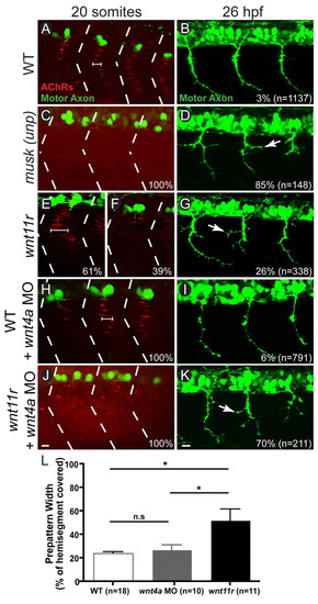
Loss of wnt11r and wnt4a phenocopies musk (unp) mutant. (A-K) Lateral view of trunk (top, dorsal; left, anterior) stained for AChR clusters (bungarotoxin, red) and motor axons (znp-1, green) in 20-somite and 26 hpf zebrafish embryos. (A,B) Three hemisegments in a wild-type embryo (hemisegment boundaries marked with dashed lines) with pre-patterned AChR clusters along the center of each hemisegment (cluster width marked by bracket; A) and three wild-type hemisegments in which motor axons have made synaptic contacts along the length of the trunk (B). (C,D) Three hemisegments in a musk (unp) mutant showing the absence of clustered AChRs, resulting in a complete dispersion of AChRs (C) and axon guidance errors in 26 hpf musk (unp) mutants (arrow points to axon branch) which occur in 85% of axons (D). (E-G) A single hemisegment from a wnt11r embryo showing an expanded AChR pre-pattern (width bracketed, 61% occurrence; E), and a single hemisegment showing a complete loss of AChR pre-pattern (39% of occurrence; F). Three hemisegments in a 26 hpf wnt11r mutant showing musk (unp)-like axon branching (arrow) in 26% of axons (G). (H,I) Three hemisegments in a wild-type embryo injected with 10 ng of wnt4a morpholino showing wild-type-like pre-patterning (H) and wild-type-like axons with only 6% guidance errors (I). (J,K) Three hemisegments from a wnt11r mutant injected with 10 ng of wnt4a morpholino showing musk (unp)-like AChR dispersal in all hemisegments (J) and guidance errors in 70% of axons (K). (L) Quantification of increase in AChR pre-pattern width in wnt11r mutants compared with wild type or wnt4a morpholino injected (P=0.01 for wild type vs wnt11r mutant, P=0.02 for wnt11r mutant vs wnt4a morpholino injected and P=0.38 for wild type vs wnt4a morpholino injected; Student’s two-tailed t-test, unequal variance; error bars indicate s.e.m.). Scale bars: 10 μm. |
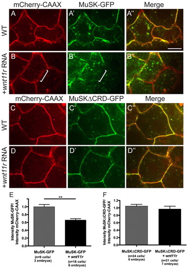
Addition of wnt11r triggers a loss of MuSK-GFP at the membrane in a CRD-dependent manner. (A-D3) Dome-stage zebrafish embryos at 4.5 hpf expressing mCherry-CAAX membrane marker (red) and either MuSK-GFP or MuSKΔCRD-GFP (green) in separate and merged channel views. (A-A3) Wild-type embryos showing MuSK-GFP membrane localization. (B-B3) Wild-type embryos injected with wnt11r mRNA showing reduced MuSK-GFP at the membrane (white bracket highlights one area of reduced GFP signal). (C-C3) Wild-type embryos showing MuSKΔCRD-GFP expression at the membrane, which is not reduced in the presence of wnt11r mRNA (D-D3). (E) Quantification of effect of wnt11r on MuSK-GFP membrane localization (**P=4.6×10⁻⁵; Student’s two-tailed t-test, unequal variance). (F) Quantification of effect of wnt11r on MuSK”CRD-GFP membrane localization (P=0.39; Student’s two-tailed t-test, unequal variance). Error bars represent s.e.m. Scale bar: 10 μm. |

MuSK-GFP localizes to Rab11-positive endosomes in the center of presynaptic muscle cells. (A) Expression of GFP driven by the smyhc1 promoter in two wild-type adaxial muscle cells (green) with pre-patterned AChR clusters (bungarotoxin, red) along the center of adaxial muscle cells. (B) Expression of 3.8musk:GFP (green) and membrane marker smyhc1:mCherry-CAAX (red) in an individual adaxial muscle cells. (C) 3.8musk:MuSK-GFP (green) and smyhc1:mCherry-CAAX (red) in a ‘young’ adaxial muscle cell that does not yet have pre-patterned AChR clusters. (D) 3.8musk:MuSK-GFP in an adaxial muscle cell (green) with AChRs (bungarotoxin, red) showing that MuSK-GFP puncta aggregate in close proximity to pre-patterned AChR clusters. (E) 3.8musk:MuSK-GFP (green) and smyhc1:mCherry-CAAX (red) in an ‘older’ adaxial muscle cell that no longer has pre-patterned AChRs, showing a reduction in central MuSK-GFP puncta (accumulation of protein at the myoseptal boundary is marked with an arrow). (F-F32) A single confocal slice showing colocalization of MuSK-RFP (red) and early/recycling endosome marker Rab11-GFP (green) expressed under the 3.8musk promoter in a normal view (F) and magnified views (F-F3). Dashed lines encircle a single muscle cell. Scale bar: 10 μm. |
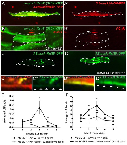
MuSK trafficking is disrupted in the absence of rab11 and wnt11r/4a. (A,A2) Co-expression of smyhc1:rab11(S25N)-GFP (green) and 3.8musk:MuSK-RFP (red; alone in A2), demonstrating disruption of MuSK-RFP center muscle localization. Instead, MuSK-RFP appears dispersed throughout the cell. (B,B2) smyhc1:rab11(S25N)-GFP (green) expressed in two adjacent muscle cells showing disruption of AChR clusters (bungarotoxin, red) at the interface of the two mosaic fibers but not in an adjacent wild-type cell (arrow). Observed in 38% of cell interfaces, n=13. (C-C3) 3.8musk:MuSK-GFP (green) in a wild-type embryo with normal protein distribution (C) and a magnified view of dashed box showing very little protein at the membrane as visualized with smyhc:mCherryCAAX (C2,C3). (D-D3) 3.8musk:MuSK-GFP (green) in a wnt11r mutant/wnt4a morphant embryo (D), and magnified view of dashed box showing accumulation of protein at the membrane as visualized with smyhc:mCherryCAAX (D2,D3). (E,F) Quantification of central enrichment of MuSK-RFP puncta when co-expressed with Rab11(S25N)-GFP (E) (P values for muscle subdivisions 1-5: 0.6, 0.27, 0.01, 0.38, 0.34, respectively), or when expressed in a wnt11r mutant/wnt4a morphant background (F; P values for muscle subdivisions 1-5: 0.87, 0.12, 0.04, 0.36, 0.97, respectively). *P<0.05. Error bars represent s.e.m. Dashed lines encircle a single muscle cell. Arrowheads (C3,D3) indicate cell membrane. Scale bars: 10 μm in A-D; 1 µm in C2-D3. |
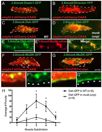
Localization of noncanonical PCP proteins to the center of muscle cells requires MuSK, and vice versa. (A,B) Daam1-GFP (green) (A) and Diversin-YFP (green) (B) under the 3.8musk promoter localize to centrally enriched puncta in muscle cells co-expressing mCherry-CAAX under the smyhc1 promoter (red). (C) Dsh-GFP under the 3.8musk promoter (green) localizes to centrally enriched puncta in fibers co-expressing mCherry-CAAX (red). (D) The punctate localization of Dsh-GFP (green) is lost in musk (unp) mutant muscle cells (mCherry-CAAX in red). (E-E3) Magnified view of center of muscle cell showing colocalization of Dsh-GFP (green) and MuSK-mKate (red), both driven by the 3.8musk promoter. (F-F3) MuSK-GFP under the 3.8musk promoter (green) localizes to centrally enriched puncta in muscle cells co-expressing mCherry-CAAX (red). F2 and F3 show magnified views of the region marked with dashed box in F. (G-G3) MuSK-GFP localizes to the membrane of muscle cells co-expressing Dsh(DEP+)-Myc. G2 and G3 show magnified views of the region marked with dashed box in G. (H) Quantification of reduction in centrally localized Dsh-GFP puncta in musk (unp) mutants (P values for muscle subdivisions 1-5: 0.49, 0.09, 0.05, 0.04, 0.02, respectively). *P<0.05. Error bars represent s.e.m. Dashed lines encircle a single muscle cell. Arrowheads in F3 and G3 indicate cell membrane. Scale bars: 10 μm. |
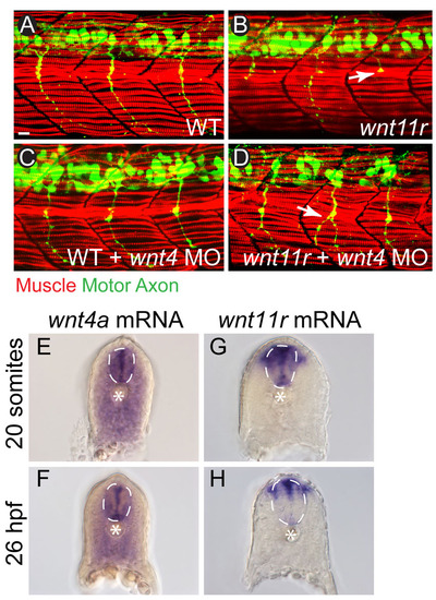
wnt4a and wnt11r are expressed in the vicinity of adaxial muscle cells and loss of Wnts does not affect muscle development. (A-D) Adaxial muscle fibers (F59, red) show normal morphology in 26 hpf wild-type embryos (A), wnt11r mutants (B), wnt4a morphants (C) and wnt11r mutant/wnt4a morphants (D). Motor axon (green) guidance is affected in wnt4a morphants (C) and wnt11r mutant/wnt4a morphants (D), as marked by white arrows. (E-H) Cross-sections of 20 somite and 26 hpf embryos stained with a DIG-labeled RNA probe for wnt4a, showing strong labeling in the spinal cord (dotted white line) as well as more diffuse staining throughout the somites. (E,F). Staining with a wnt11r probe revealed strong labeling in the spinal cord as well as a narrow region of dorsolateral somatic staining lateral to the spinal cord (G,H). Notochord is marked with an asterisk. |
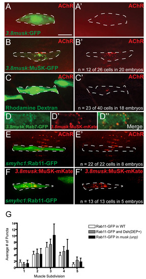
3.8musk:MuSK-GFP rescues AChR clustering to the same degree as transplanting wild-type cells into musk mutants, and distribution of Rab11-GFP and Rab7-GFP in muscle cells. (A,A2) Control expression of 3.8musk:GFP (green) in musk(unp) mutants fails to rescue AChR pre-patterning (bungarotoxin, red) (merged channels in A and red alone in A2). (B,B2) Expression of 3.8musk:MuSK-GFP in unplugged mutants at the 20-somite stage rescues AChR clusters (bungarotoxin, red) cell-autonomously in 47% of embryos (n=12 of 26 muscle cells in 20 embryos; merged channels in B, red alone in B2). (C,C2) 20-somite-stage wild-type derived adaxial muscle cell labeled with rhodamine dextran (green; transplanted at blastula stage) in an otherwise musk(unp) mutant embryo rescues AChR clusters (bungarotoxin, red) in 57% of embryos (n=23 of 40 cells in 18 embryos; merged channels in C, red alone in C2). (D-D22) Magnified view of the center of a 20-somite-stage muscle fiber showing weak colocalization of Rab7-GFP (green, green channel alone in D) and MuSK-mKate (red, red channel alone in D2) under the promoter (merged channels in D22). (E-F2) High level expression of rab11-GFP (green) via expression of smyhc1:rab11-GFP in individual, wild-type 20-somite-stage muscle cells does not alter AChR localization (bungarotoxin, red; n=22 of 22 cells in eight embryos; E,E2) or MuSK-mKate localization (n=13 of 13 cells in five embryos; F-F2). (G) Quantification of the distribution of Rab11-GFP puncta in wild-type muscle cells, musk mutant muscle cells or in cells co-expressing Dsh(DEP+)-myc. Scale bar: 10 μm. |
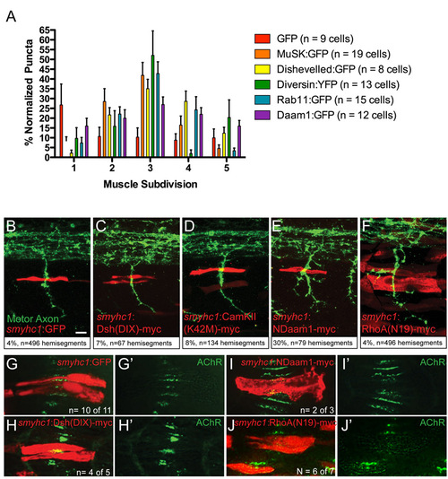
Expression of dominant-negative PCP but not canonical Wnt pathway components in muscle cells disrupts AChR clustering and axon guidance. (A) Quantification of the distribution of puncta of various proteins under the control of the 3.8musk promoter among five laterally delineated segments of muscle cells, normalized to 100. (B-F) Stochastic expression of smyhc1:GFP (B), smyhc1:dsh(DIX)-myc (C) and smyhc1: camKII(K42M)-myc (D) (all in red) in muscle cells just dorsal to the horizontal myoseptum in 27 hpf embryos does not induce axon guidance errors (axons in green). Expression of smyhc1:NDaam1-myc (E) and smyhc1:rhoA(N19)-myc (F) (both in red) does induce guidance errors (white arrow, motor axons stained with znp-1 in green). Axon guidance defects in %. (G-J2) Stochastic expression of smyhc1:Ndaam1-myc (I,I2) and smyhc1:rhoA(N19)-myc (J,J2) but not smyhc1:GFP (G,G2) or smyhc1:dsh(DIX)-myc (H,H2) (all in red) in 20-somite wild-type embryos causes a reduction in AChR pre-patterned clustering (bungarotoxin, red) at the interface between the two expressing fibers but not in adjacent wild-type fibers. Scale bar; 10 μm. |