
|
Fig. 3
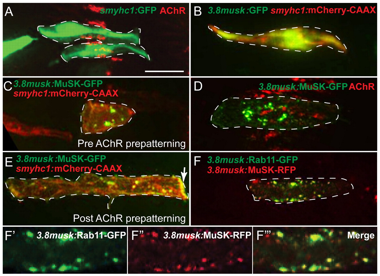
MuSK-GFP localizes to Rab11-positive endosomes in the center of presynaptic muscle cells. (A) Expression of GFP driven by the smyhc1 promoter in two wild-type adaxial muscle cells (green) with pre-patterned AChR clusters (bungarotoxin, red) along the center of adaxial muscle cells. (B) Expression of 3.8musk:GFP (green) and membrane marker smyhc1:mCherry-CAAX (red) in an individual adaxial muscle cells. (C) 3.8musk:MuSK-GFP (green) and smyhc1:mCherry-CAAX (red) in a ‘young’ adaxial muscle cell that does not yet have pre-patterned AChR clusters. (D) 3.8musk:MuSK-GFP in an adaxial muscle cell (green) with AChRs (bungarotoxin, red) showing that MuSK-GFP puncta aggregate in close proximity to pre-patterned AChR clusters. (E) 3.8musk:MuSK-GFP (green) and smyhc1:mCherry-CAAX (red) in an ‘older’ adaxial muscle cell that no longer has pre-patterned AChRs, showing a reduction in central MuSK-GFP puncta (accumulation of protein at the myoseptal boundary is marked with an arrow). (F-F32) A single confocal slice showing colocalization of MuSK-RFP (red) and early/recycling endosome marker Rab11-GFP (green) expressed under the 3.8musk promoter in a normal view (F) and magnified views (F-F3). Dashed lines encircle a single muscle cell. Scale bar: 10 μm.