
Fig. 2
- ID
- ZDB-FIG-120217-18
- Publication
- Gordon et al., 2012 - Initiation of synapse formation by Wnt-induced MuSK endocytosis
- Other Figures
- All Figure Page
- Back to All Figure Page
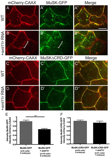
Addition of wnt11r triggers a loss of MuSK-GFP at the membrane in a CRD-dependent manner. (A-D3) Dome-stage zebrafish embryos at 4.5 hpf expressing mCherry-CAAX membrane marker (red) and either MuSK-GFP or MuSKΔCRD-GFP (green) in separate and merged channel views. (A-A3) Wild-type embryos showing MuSK-GFP membrane localization. (B-B3) Wild-type embryos injected with wnt11r mRNA showing reduced MuSK-GFP at the membrane (white bracket highlights one area of reduced GFP signal). (C-C3) Wild-type embryos showing MuSKΔCRD-GFP expression at the membrane, which is not reduced in the presence of wnt11r mRNA (D-D3). (E) Quantification of effect of wnt11r on MuSK-GFP membrane localization (**P=4.6×10⁻⁵; Student’s two-tailed t-test, unequal variance). (F) Quantification of effect of wnt11r on MuSK”CRD-GFP membrane localization (P=0.39; Student’s two-tailed t-test, unequal variance). Error bars represent s.e.m. Scale bar: 10 μm. |