
Fig. S1
- ID
- ZDB-FIG-120217-22
- Publication
- Gordon et al., 2012 - Initiation of synapse formation by Wnt-induced MuSK endocytosis
- Other Figures
- All Figure Page
- Back to All Figure Page
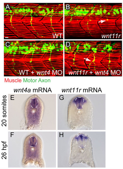
wnt4a and wnt11r are expressed in the vicinity of adaxial muscle cells and loss of Wnts does not affect muscle development. (A-D) Adaxial muscle fibers (F59, red) show normal morphology in 26 hpf wild-type embryos (A), wnt11r mutants (B), wnt4a morphants (C) and wnt11r mutant/wnt4a morphants (D). Motor axon (green) guidance is affected in wnt4a morphants (C) and wnt11r mutant/wnt4a morphants (D), as marked by white arrows. (E-H) Cross-sections of 20 somite and 26 hpf embryos stained with a DIG-labeled RNA probe for wnt4a, showing strong labeling in the spinal cord (dotted white line) as well as more diffuse staining throughout the somites. (E,F). Staining with a wnt11r probe revealed strong labeling in the spinal cord as well as a narrow region of dorsolateral somatic staining lateral to the spinal cord (G,H). Notochord is marked with an asterisk. |