
|
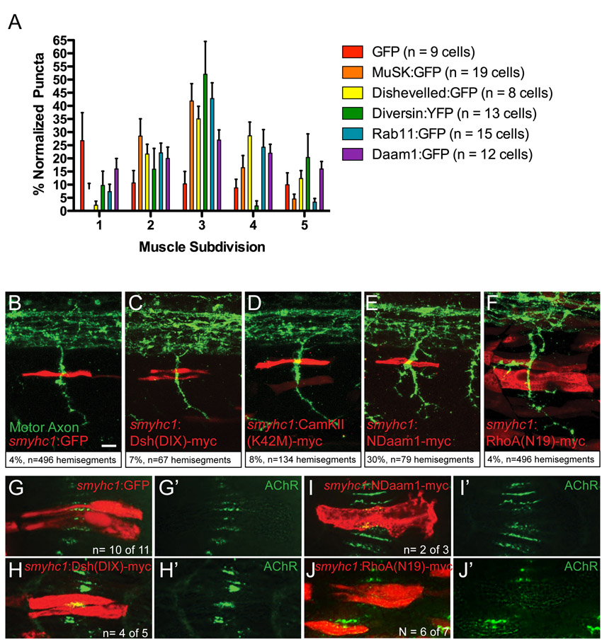
Fig. S3
Expression of dominant-negative PCP but not canonical Wnt pathway components in muscle cells disrupts AChR clustering and axon guidance. (A) Quantification of the distribution of puncta of various proteins under the control of the 3.8musk promoter among five laterally delineated segments of muscle cells, normalized to 100. (B-F) Stochastic expression of smyhc1:GFP (B), smyhc1:dsh(DIX)-myc (C) and smyhc1: camKII(K42M)-myc (D) (all in red) in muscle cells just dorsal to the horizontal myoseptum in 27 hpf embryos does not induce axon guidance errors (axons in green). Expression of smyhc1:NDaam1-myc (E) and smyhc1:rhoA(N19)-myc (F) (both in red) does induce guidance errors (white arrow, motor axons stained with znp-1 in green). Axon guidance defects in %. (G-J2) Stochastic expression of smyhc1:Ndaam1-myc (I,I2) and smyhc1:rhoA(N19)-myc (J,J2) but not smyhc1:GFP (G,G2) or smyhc1:dsh(DIX)-myc (H,H2) (all in red) in 20-somite wild-type embryos causes a reduction in AChR pre-patterned clustering (bungarotoxin, red) at the interface between the two expressing fibers but not in adjacent wild-type fibers. Scale bar; 10 μm.