
- Title
-
Analysis of leukemia inhibitory factor and leukemia inhibitory factor receptor in embryonic and adult zebrafish (Danio rerio)
- Authors
- Hanington, P.C., Patten, S.A., Reaume, L.M., Waskiewicz, A.J., Belosevic, M., and Ali, D.W.
- Source
- Full text @ Dev. Biol.
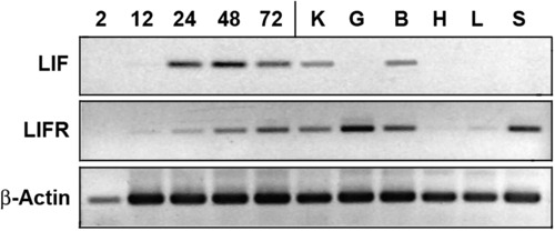
RT–PCR showing expression of lif and lifr in zebrafish embryos 2, 12, 24, 48 and 72 h post fertilization, and in the kidney (K), gill (G), brain (B), heart (H), liver (L) and spleen (S) of adult zebrafish. lif and lifr expression was compared to a β-actin loading control. |
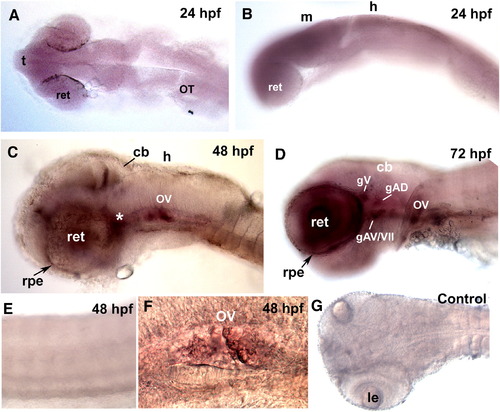
Embryonic and larval expression patterns of lif. (A, B) lif expression was observed to be most prominent in the retina, however expression was widespread and there was no exclusive expression of lif in distinct areas of the brain. (C) At 48 hpf, lif was clearly visible in regions of the cranial sensory ganglia (asterisk, *), otic vesicle (OV), retina (ret) and area of the midbrain–hindbrain boundary. (D) At 72 hpf, lif was present at relatively high levels in the retina, otic vesicle and the cranial sensory ganglia. lif was localized in the presumptive trigeminal (gV), facial (gVII), anterioventral ganglion (gAV) and anteriodorsal ganglion (gAD). (E) lif was not expressed in muscle and trunk neurons. (F) Expression of lif in the otic vesicle was visible at both 48 hpf and 72 hpf. (G) In situ hybridization pattern of the lif sense probe. (t), telencephalon; (m), midbrain; (h), hindbrain; (cb), cerebellum; (rpe), retinal pigmented epithelium; (le), lens. EXPRESSION / LABELING:
|
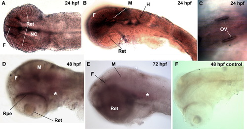
Embryonic and larval expression patterns of lifr. (A, B) At 24 hpf, lifr was highly expressed in the forebrain, midbrain and notochord. (C) lifr was also visible in the otic vesicle (OV) at 24 hpf. (D) At 48 hpf, lifr was localized in regions of the cranial sensory ganglia (asterisk, *), retina and forebrain. (E) Expression pattern of lifr at 72 hpf was similar to that at 48 hpf. lifr was expressed in regions of the cranial sensory ganglia (*) and forebrain. (F) lifr was not expressed in trunk muscle and trunk neurons. (f), forebrain; (m), midbrain; (h), hindbrain; (cb), cerebellum; (rpe), retinal pigmented epithelium. EXPRESSION / LABELING:
PHENOTYPE:
|
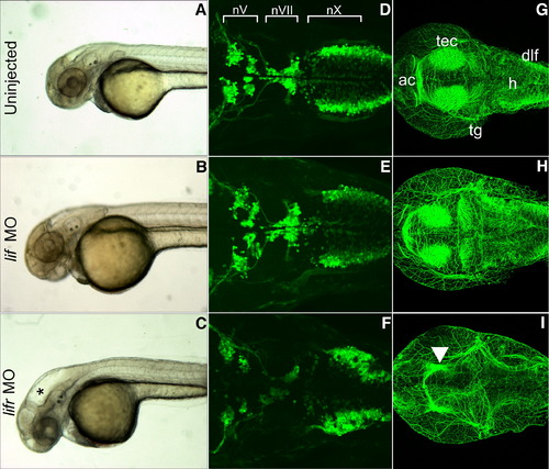
Lifr-depleted, but not Lif-depleted embryos exhibit hydrocephaly and neural defects. (A–C) Live images of 48 hpf embryos in lateral view with anterior to left. Embryos are wild type uninjected (A), injected with lif MO (B), or injected with lifr MO (C). Asterisk (*) marks hydrocephaly observed in lifr morphants, but not in lif MO-injected or uninjected embryos. (D–F) Confocal fluorescent composite images of hindbrain branchiomotor neurons in wild type uninjected (D), lif MO-injected (E), or lifr MO-injected (F) 48 hpf Isl1-GFP transgenic embryos. View is dorsal with anterior to left. (G–I) Confocal fluorescent composite images showing anti-acetylated antibody-stained cranial axons of 48 hpf embryos. View is dorsal with anterior to left. Embryos are wild type uninjected (G), lif MO-injected (H), or lifr MO-injected (I). Arrowhead indicates significant loss of staining in the tectum of lifr morphants. (tec), tectum; (ac), anterior commissure neurons; (dlf), dorsal longitudinal fasciculus; (h), hindbrain; (tg), trigeminal ganglion. |
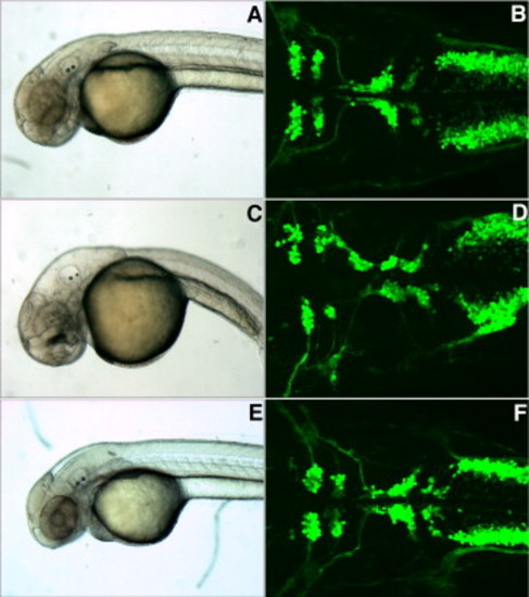
Rescue of the lifr MO phenotype by co-injection of the lifr MO with lifr synthetic mRNA. Uninjected embryos exhibited no morphological defects (A) and developed normal cranial ganglia (B) at 48 hpf. In contrast, lifr MO injected embryos exhibited severe hydrocephaly (C) and possess several defects in branchiomotor neuron development (D) at 48 hpf. Co-injection of lifr MO with lifr mRNA rescued the lifr MO phenotype and resulted in ∼92% of embryos displaying a normal morphological phenotype (E), and ∼97% of embryos developing normal branchiomotor neurons (F). |
Reprinted from Developmental Biology, 314(2), Hanington, P.C., Patten, S.A., Reaume, L.M., Waskiewicz, A.J., Belosevic, M., and Ali, D.W., Analysis of leukemia inhibitory factor and leukemia inhibitory factor receptor in embryonic and adult zebrafish (Danio rerio), 250-260, Copyright (2008) with permission from Elsevier. Full text @ Dev. Biol.