
Fig. 4
- ID
- ZDB-FIG-080331-20
- Publication
- Hanington et al., 2008 - Analysis of leukemia inhibitory factor and leukemia inhibitory factor receptor in embryonic and adult zebrafish (Danio rerio)
- Other Figures
- All Figure Page
- Back to All Figure Page
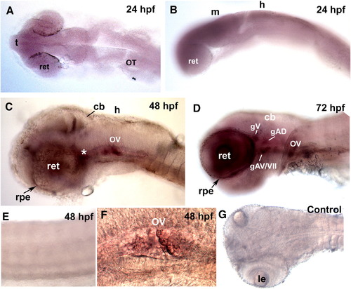
Embryonic and larval expression patterns of lif. (A, B) lif expression was observed to be most prominent in the retina, however expression was widespread and there was no exclusive expression of lif in distinct areas of the brain. (C) At 48 hpf, lif was clearly visible in regions of the cranial sensory ganglia (asterisk, *), otic vesicle (OV), retina (ret) and area of the midbrain–hindbrain boundary. (D) At 72 hpf, lif was present at relatively high levels in the retina, otic vesicle and the cranial sensory ganglia. lif was localized in the presumptive trigeminal (gV), facial (gVII), anterioventral ganglion (gAV) and anteriodorsal ganglion (gAD). (E) lif was not expressed in muscle and trunk neurons. (F) Expression of lif in the otic vesicle was visible at both 48 hpf and 72 hpf. (G) In situ hybridization pattern of the lif sense probe. (t), telencephalon; (m), midbrain; (h), hindbrain; (cb), cerebellum; (rpe), retinal pigmented epithelium; (le), lens. |
| Gene: | |
|---|---|
| Fish: | |
| Anatomical Terms: | |
| Stage Range: | Prim-5 to Protruding-mouth |
Reprinted from Developmental Biology, 314(2), Hanington, P.C., Patten, S.A., Reaume, L.M., Waskiewicz, A.J., Belosevic, M., and Ali, D.W., Analysis of leukemia inhibitory factor and leukemia inhibitory factor receptor in embryonic and adult zebrafish (Danio rerio), 250-260, Copyright (2008) with permission from Elsevier. Full text @ Dev. Biol.