
Fig. 6
- ID
- ZDB-FIG-080331-22
- Publication
- Hanington et al., 2008 - Analysis of leukemia inhibitory factor and leukemia inhibitory factor receptor in embryonic and adult zebrafish (Danio rerio)
- Other Figures
- All Figure Page
- Back to All Figure Page
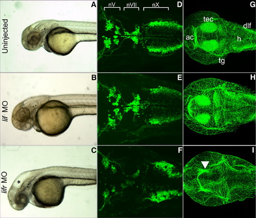
Lifr-depleted, but not Lif-depleted embryos exhibit hydrocephaly and neural defects. (A–C) Live images of 48 hpf embryos in lateral view with anterior to left. Embryos are wild type uninjected (A), injected with lif MO (B), or injected with lifr MO (C). Asterisk (*) marks hydrocephaly observed in lifr morphants, but not in lif MO-injected or uninjected embryos. (D–F) Confocal fluorescent composite images of hindbrain branchiomotor neurons in wild type uninjected (D), lif MO-injected (E), or lifr MO-injected (F) 48 hpf Isl1-GFP transgenic embryos. View is dorsal with anterior to left. (G–I) Confocal fluorescent composite images showing anti-acetylated antibody-stained cranial axons of 48 hpf embryos. View is dorsal with anterior to left. Embryos are wild type uninjected (G), lif MO-injected (H), or lifr MO-injected (I). Arrowhead indicates significant loss of staining in the tectum of lifr morphants. (tec), tectum; (ac), anterior commissure neurons; (dlf), dorsal longitudinal fasciculus; (h), hindbrain; (tg), trigeminal ganglion. |
Reprinted from Developmental Biology, 314(2), Hanington, P.C., Patten, S.A., Reaume, L.M., Waskiewicz, A.J., Belosevic, M., and Ali, D.W., Analysis of leukemia inhibitory factor and leukemia inhibitory factor receptor in embryonic and adult zebrafish (Danio rerio), 250-260, Copyright (2008) with permission from Elsevier. Full text @ Dev. Biol.