
|
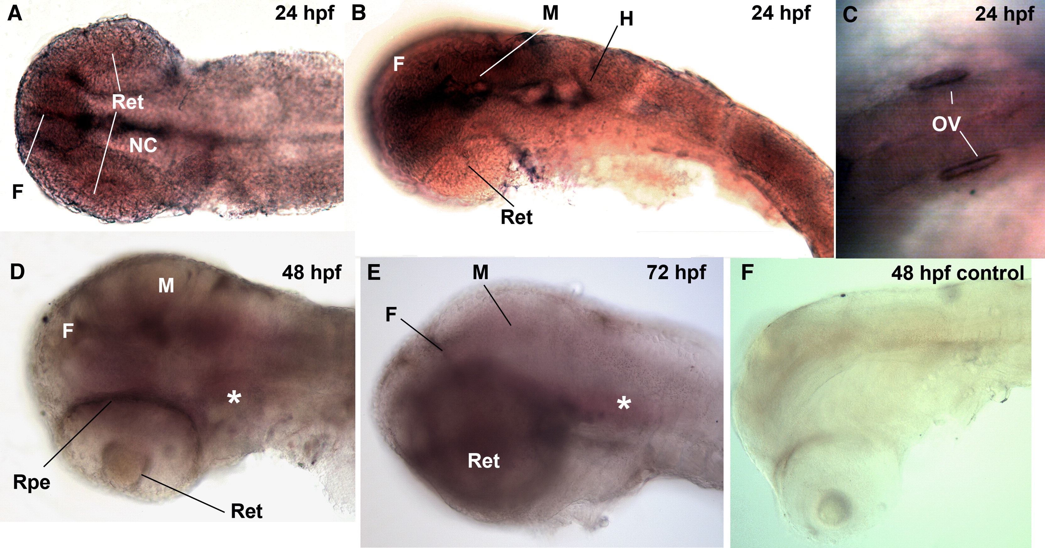
Fig. 5 Embryonic and larval expression patterns of lifr. (A, B) At 24 hpf, lifr was highly expressed in the forebrain, midbrain and notochord. (C) lifr was also visible in the otic vesicle (OV) at 24 hpf. (D) At 48 hpf, lifr was localized in regions of the cranial sensory ganglia (asterisk, *), retina and forebrain. (E) Expression pattern of lifr at 72 hpf was similar to that at 48 hpf. lifr was expressed in regions of the cranial sensory ganglia (*) and forebrain. (F) lifr was not expressed in trunk muscle and trunk neurons. (f), forebrain; (m), midbrain; (h), hindbrain; (cb), cerebellum; (rpe), retinal pigmented epithelium.
Reprinted from Developmental Biology, 314(2), Hanington, P.C., Patten, S.A., Reaume, L.M., Waskiewicz, A.J., Belosevic, M., and Ali, D.W., Analysis of leukemia inhibitory factor and leukemia inhibitory factor receptor in embryonic and adult zebrafish (Danio rerio), 250-260, Copyright (2008) with permission from Elsevier. Full text @ Dev. Biol.