
- Title
-
Neuropilin asymmetry mediates a left-right difference in habenular connectivity
- Authors
- Kuan, Y.S., Yu, H.H., Moens, C.B., and Halpern, M.E.
- Source
- Full text @ Development
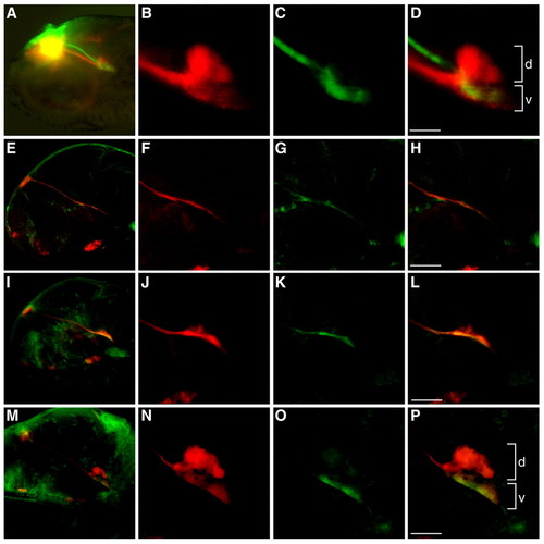
Differential dorsoventral innervation of the IPN by the left and right habenulae. (A) Dorsolaterial view of the habenulo-IPN projections in a 4-day-old zebrafish larva. DiI (red) and DiO (green) were injected into the left and right habenula, respectively. The yellow signal is a visual artifact owing to the superimposition of the differentially-labeled L-R habenulae in this orientation. (B-D) Higher magnification of A confirms that the left habenula innervates dorsal (d) and ventral (v) IPN, whereas the right habenula only projects ventrally. (E-P) Lateral views of (E-H) 2-, (I-L) 3- and (M-P) 4-day-old larvae labeled with anti-Lov (red) and anti-Ron (green) antibodies. F-H, J-L and N-P are higher magnifications of E, I and M, respectively. Scale bars: 50 μm. |
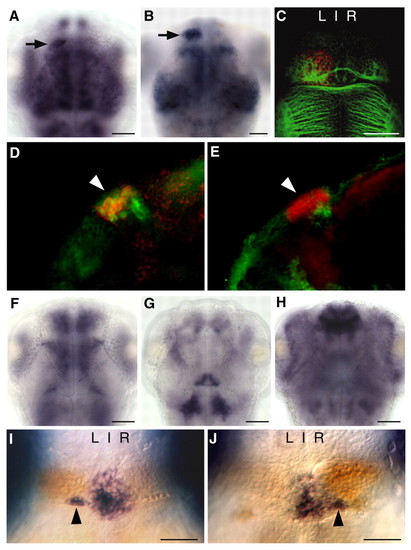
Asymmetric expression of nrp1a in the dorsal diencephalon. (A,B) Left-sided expression of nrp1a (black arrow) is detected by 2 days (A) and increases by 4 days (B). (C) nrp1a expression (red) overlaps with the dense neuropil of the left habenula revealed by anti-acetylated tubulin immunolabeling (green) at 4 days. (D,E) The nrp1a-expressing neurons (red) are a subset of the Lov-immunoreactive population (D, green) and are also distinct from Ron-immunoreactive cells (E, green) of the left habenula (arrowheads). (F-H) Expression of (F) nrp1b, (G) nrp2a and (H) nrp2b was not detected in the habenular nuclei. (I,J) Laterality of nrp1a expression (orange) correlates with parapineal position (otx5 expression, black arrowheads) in (I) mock-injected (94±2% left bias, n=89) and (J) spaw MO-injected 4-day-old larvae (56±2% sinistral and 44±2% dextral, n=96). A-C and F-J are dorsal views of the brain; C and D are lateral views of composite Z-stack confocal images through the left habenula. Left (L) and right (R) sides of the brain are indicated. Scale bars: 50 μm. |
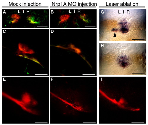
Innervation of the dorsal IPN requires Nrp1a. Dorsal views of habenular nuclei (A,B,G,H) and lateral views of the IPN (C,D,E,F,I) of 4-day-old larvae. (A-D) Mock-injected (A,C) or Nrp1a MO-injected (B,D) larvae were labeled with anti-Lov (red) and anti-Ron antibody (green). (E,F,I) Mock-injected (E), Nrp1a MO-injected (F) or parapineal-ablated (I) larvae were labeled at 4 days with DiI (red) in the left habenula. (G) All control-ablated larvae (n=27) had an intact parapineal (black arrowhead) as confirmed by otx5 expression (blue), and strong nrp1a expression (orange) in the left habenula. (H) In the majority of parapineal-ablated larvae, nrp1a expression (orange) was greatly reduced in the left habenula (∼71%, n=39). Scale bars: 50 μm. |
Innervation of the dorsal IPN requires Nrp1a. Dorsal views of habenular nuclei (A,B,G,H) and lateral views of the IPN (C,D,E,F,I) of 4-day-old larvae. (A-D) Mock-injected (A,C) or Nrp1a MO-injected (B,D) larvae were labeled with anti-Lov (red) and anti-Ron antibody (green). (E,F,I) Mock-injected (E), Nrp1a MO-injected (F) or parapineal-ablated (I) larvae were labeled at 4 days with DiI (red) in the left habenula. (G) All control-ablated larvae (n=27) had an intact parapineal (black arrowhead) as confirmed by otx5 expression (blue), and strong nrp1a expression (orange) in the left habenula. (H) In the majority of parapineal-ablated larvae, nrp1a expression (orange) was greatly reduced in the left habenula (∼71%, n=39). Scale bars: 50 μm. |
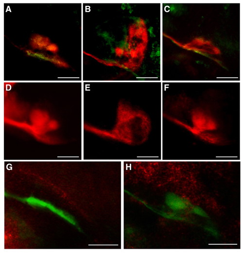
Overexpression of sema3D causes ectopic left habenular projections. (A-F) Lateral views of the 4-day-old larval IPN from (A,D) mock-injected embryos (n=91) or embryos injected with (B,E) sema3D mRNA (n=64), or (C,F) sema3Gb mRNA (n=69). Larvae were stained with anti-Lov antibody (red) and anti-Ron antibody (green) in A-C, or labeled with DiI (red) in the left habenula in D-F. (G,H) Lateral views of (G) 2- and (H) 4-day-old larvae double-labeled with sema3D antisense RNA probe and anti-Lov antibody. sema3d transcripts (red) are abundant in the brain region dorsal to the IPN, as visualized by Lov+ habenular projections (green). Scale bars: 50 μm. |
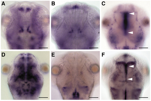
Expression patterns of semaphorin-related genes in the larval brain. Dorsal views at 2 days of (A) sema3Aa, (B) sema3Ab, (C) sema3D, (D) sema3Fa, (E) sema3Ga and (F) sema3Gb expression. Arrowheads indicate expression in the vicinity of the FR tracts from the diencephalon to the midbrain. Scale bars: 50 μm. |