|
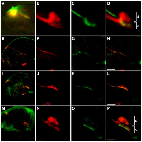
Differential dorsoventral innervation of the IPN by the left and right habenulae. (A) Dorsolaterial view of the habenulo-IPN projections in a 4-day-old zebrafish larva. DiI (red) and DiO (green) were injected into the left and right habenula, respectively. The yellow signal is a visual artifact owing to the superimposition of the differentially-labeled L-R habenulae in this orientation. (B-D) Higher magnification of A confirms that the left habenula innervates dorsal (d) and ventral (v) IPN, whereas the right habenula only projects ventrally. (E-P) Lateral views of (E-H) 2-, (I-L) 3- and (M-P) 4-day-old larvae labeled with anti-Lov (red) and anti-Ron (green) antibodies. F-H, J-L and N-P are higher magnifications of E, I and M, respectively. Scale bars: 50 μm.
|