
- Title
-
Cloning of full-length zebrafish dcc and expression analysis during embryonic and early larval development
- Authors
- Fricke, C., and Chien, C.B.
- Source
- Full text @ Dev. Dyn.
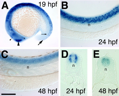
A-E: Expression analysis of dcc in the spinal cord (SC) in whole-mounts (A-C) and transverse sections (D,E). A-C: Expression in the SC was at first patchy (A, 19 hours postfertilization [hpf] and B, 24 hpf) but became more uniform later in development (C, 48 hpf). D,E: Expression was first laterally restricted (D, 24 hpf) then spread more medially (E, 48 hpf). Note that the expression in the dorsal SC (thick arrow in A) reached further posterior than that in ventral SC (small arrow in A) at 19 hpf. Expression was also found in the epiphysis (small arrowhead in A), olfactory placode and bulb (large arrowhead in A), and in the ventrorostral neuronal cluster of the forebrain (+ in A) at 19 hpf. By 22 hpf, specific expression was also detected in the ventrocaudal neuronal cluster (not shown). Lateral views in A-C; transverse sections in D,E. Rostral is to the left, dorsal up in B and C. n, notochord. Scale bar = 200 μm in A, 100 μm in B,C, 50 μm in D,E. EXPRESSION / LABELING:
|

A-C: dcc is expressed in the developing hindbrain at 22 hours postfertilization (hpf; A), 24 hpf (B), and 48 hpf (C). The number of cells in the hindbrain expressing dcc increased during development. Regions of denser expression persist at later stages (asterisks). D: Double staining with dcc and krox-20 (+ in D) at 18 hpf. krox-20 labels the third and fifth rhombomeres. Dorsal views, rostral to the left. r, rostral; c, caudal. Scale bar = 100 μm. EXPRESSION / LABELING:
|
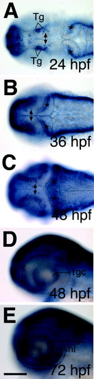
dcc is expressed dynamically in the visual system. A-C: dcc in the tectum was first detected at 24 hours postfertilization (hpf) in a few cells (double arrow in A), had spread by 36 hpf (double arrow in B), and became restricted to cells located at the tectal margin by 48 hpf (double arrow in C). dcc expression was also found in the tegmentum (A, Tg) and cerebellum (stars in A-C). D,E: In the eye, dcc mRNA is expressed in cells of the retinal ganglion cell (RGC) layer (D, E, 48 and 72 hpf, respectively) and later also in cells of the inner nuclear layer (INL; E, 72 hpf). Expression in both layers was detected up to 120 hpf, the latest time point analyzed (not shown). Dorsal views in A-C; lateral views in D,E. Rostral is to the left. rgc, retinal ganglion cell layer; inl, inner nuclear layer. Scale bar = 100 μm in A-E. EXPRESSION / LABELING:
|
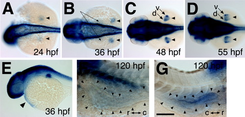
dcc expression in non-neural tissues. A-D: Whereas dcc was found to be expressed uniformly in the mesenchyme of the fin bud at 24 and 36 hours postfertilization (hpf; arrowheads in A,B), it became more strongly expressed in the dorsal (d in C,D) and ventral (v in C,D) regions at 48 (C) and 55 (D) hpf. dcc was also expressed by cells located on the yolk sac anterior and medial to the fin bud (long arrows in B), presumably corresponding to the developing pronephros. E-G: Furthermore, dcc was expressed in the heart (arrowhead in E, 36 hpf), anterior to mid-intestine (arrowheads in F, 120 hpf, left side view), and pancreas (arrowheads in G, 120 hpf, right side view) during later stages of development. Dorsal views in A-D; lateral views in E-G. r, rostral; c, caudal. Scale bars = 200 μm in A-E, 100 μm in F,G. |

Unillustrated author statements EXPRESSION / LABELING:
|