|
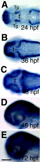
dcc is expressed dynamically in the visual system. A-C: dcc in the tectum was first detected at 24 hours postfertilization (hpf) in a few cells (double arrow in A), had spread by 36 hpf (double arrow in B), and became restricted to cells located at the tectal margin by 48 hpf (double arrow in C). dcc expression was also found in the tegmentum (A, Tg) and cerebellum (stars in A-C). D,E: In the eye, dcc mRNA is expressed in cells of the retinal ganglion cell (RGC) layer (D, E, 48 and 72 hpf, respectively) and later also in cells of the inner nuclear layer (INL; E, 72 hpf). Expression in both layers was detected up to 120 hpf, the latest time point analyzed (not shown). Dorsal views in A-C; lateral views in D,E. Rostral is to the left. rgc, retinal ganglion cell layer; inl, inner nuclear layer. Scale bar = 100 μm in A-E.
|