
- Title
-
Zebrafish Lmx1b.1 and Lmx1b.2 are required for maintenance of the isthmic organizer
- Authors
- O'Hara, F.P., Beck, E., Barr, L.K., Wong, L.L., Kessler, D.S., and Riddle, R.D.
- Source
- Full text @ Development
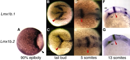
Expression of lmx1b.1 and lmx1b.2 in wild-type embryos. (A) Lateral view of a gastrula at 90% epiboly, animal pole leftwards. lmx1b.2 is expressed in the blastoderm margin (black arrowhead). (B-E) Dorsal views with anterior towards the left. (B,C) At the tailbud stage, lmx1b.1 and lmx1b.2 are both expressed in chevron-shaped domains at the presumptive MMR. Both are also expressed in the midline, with lmx1b.1 extending more rostrally and lmx1b.2 extending more caudally. (D,E) By the five-somite stage, expression of lmx1b.1 and lmx1b.2 are refined to rings at the MMR. lmx1b.1 is now expressed at the developing otic placode, while lmx1b.2 is expressed in the diencephalic region and in cells of the hindbrain that are converging on the dorsal midline. (F,G) Lateral views, rostral leftwards. By the 13-somite stage, lmx1b.1 and lmx1b.2 are both stably expressed at the MMR, the ventral diencephalon and the dorsal midline of the caudal CNS. lmx1b.1 is also stably expressed in the otic placodes. Red arrowheads indicate the MMR. ov, otic vesicle; dd, dorsal diencephalon; vd, ventral diencephalon.
EXPRESSION / LABELING:
|
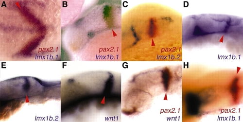
lmx1b.1, lmx1b.2, wnt1 and pax2.1 are expressed in overlapping domains at the isthmus. (A) Dorsal view, with anterior leftwards. At the one-somite stage, lmx1b.1 is expressed in the midline and in the precursors of the MMR and the otic placodes. At the presumptive IsO, pax 2.1 expression completely overlaps with lmx1b.1 expression and the pax2.1 domain is slightly broader. (B-G) Lateral views, with dorsal upwards and anterior leftwards. (B,C) At the 13-somite stage, pax2.1 expression at the IsO overlaps the expression of lmx1b.1 (B) and lmx1b.2 (C), and extends posteriorly. (D-F) At 24 hpf, the expression domains of lmx1b.1 (D), lmx1b.2 (E) and wnt1 (F) are found in the posterior mesencephalic vesicle, abutting the metencephalic vesicle. (G,H)At 24 hpf, pax2.1 expression overlaps both wnt1 (G) and lmx1b.1 (H, dorsolateral view), and extends more posteriorly. Red arrowheads indicate the MMR.
EXPRESSION / LABELING:
|
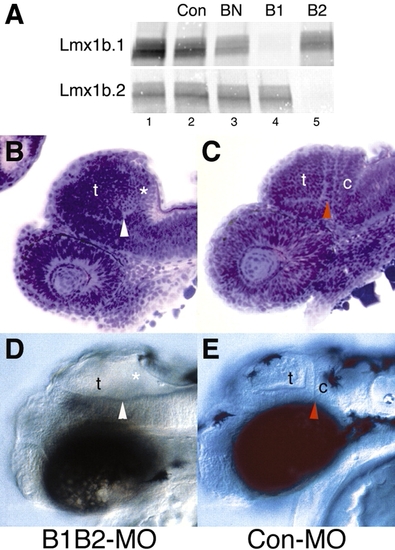
Simultaneous knockdown of Lmx1b.1 and Lmx1b.2 results in a loss of isthmic and cerebellar structures. (A) Specific inhibition of Lmx1b.1 and Lmx1b.2 translation by morpholino oligonucloetides. In vitro translation of Lmx1b.1 (top panel) and Lmx1b.2 (bottom panel) in the absence of oligonuclotide (lane 1) or with addition of: an unrelated oligonucleotide (CON-MO, lane 2), a mismatch oligonucleotide (BN-MO lane 3), or oligonucleotides specific for Lmx1b.1 (B1-MO, lane 4) or Lmx1b.2 (B2-MO, lane 5). (B-E) Lateral views of anterior CNS, with dorsal upwards and anterior leftwards. Microtome sections at 30 hpf (B,C) and whole mounts at 36 hpf (D,E) of embryos injected with B1B2-MO (B,D) or Con-MO (C,E). Red arrowheads indicate the position of the isthmic constriction and white arrowheads indicate the missing isthmic constriction. t, tectum; c, cerebellum; *, missing structures.
|
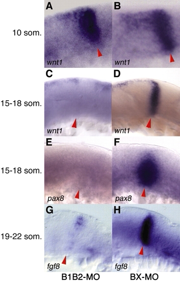
Following knockdown of Lmx1b.1 and Lmx1b.2, IsO genes are initiated normally, but some are not maintained. Lateral views of anterior CNS, with dorsal upwards and anterior leftwards. Embryos were injected with B1B2-MO (A,C,E,G) or BX-MO (B,D,F,H). wnt1 is initiated normally at the 10-somite stage. (A,B), but fades at the MMR by 15-18 somites (C,D). pax8 expression fades by 15-18 somites (E,F), and fgf8 fades by 19-22 somites, with partial dorsal retention (G,H). Red arrowheads indicate the MMR.
EXPRESSION / LABELING:
|
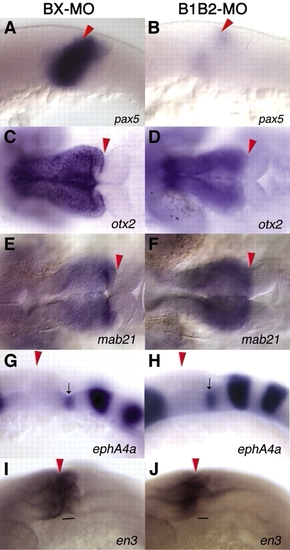
Knockdown of Lmx1b.1 and Lmx1b.2 affects cell fate in the isthmocerebellar region, but not the mesencephalon or ventral rhombomere 1 (R1). Lateral (A,B,G,H), dorsal (C-F) or dorsolateral views (I,J), with anterior leftwards. Embryos were injected with BX-MO (A,C,E,G,I) or B1B2-MO (B,D,F,H,J). pax5 expression (A,B), which marks the isthmus and cerebellum, is strongly reduced at 24 hpf in Lmx1b.1/2 knockdown embryos. Expression of otx2 (C,D) and mab21 (E,F), markers of the mesencephalon, is unaffected Lmx1b.1/2 knockdown embryos. Epha4a expression (G,H) marks several regions of the developing brain, including the ventral region of R1, and is unaffected in Lmx1b.1/2 knockdown embryos. Expression of en3 (I,J) marks both the tectum and the isthmocerebellar region. Knockdown of Lmx1b.1/2 results in a loss of en3 expression at the ventral isthmus, but normal expression is retained in the tectum and dorsal isthmus. Red arrowheads indicate the position of the MMR, black arrows indicate the ventral R1 staining of epha4a and horizontal lines indicate the ventral isthmic staining of en3.
EXPRESSION / LABELING:
|
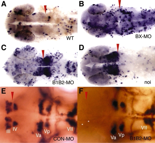
Knockdown of Lmx1b.1 and Lmx1b.2 results in increased cell death around the IsO, with a concomitant failure of neural differentiation. (A-D) A TUNEL assay stains apoptotic cells blue. Dorsal views with anterior leftwards. (A) A low level of randomly distributed apoptosis is detected in wild-type embryos. (B) Injection of BX-MO results in a nonspecific increase in apoptosis. (C) Injection of B1B2-MO sharply increases apoptosis in the IsO, especially caudally. Nonspecific apoptosis is also increased. (D) Increased apoptosis is detected around the IsO in embryos homozygous for the pax2.1/no isthmus mutation, especially rostrally. (E,F) Cranial motoneurons were visualized by expressing GFP under the control of the Islet1 promoter and staining with an antibody against GFP. (E) Cranial motoneurons III and IV flank the IsO. (F) Neurons III and IV fail to form in B1B2-MO-injected embryos, while other neurons are unaffected. Red arrowheads indicate position of the MMR and asterisks mark normal position of missing neurons.
|
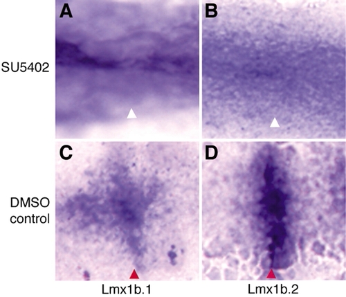
Blocking of FGF receptor activity results in loss of lmx1b.1 and lmx1b.2 expression at the developing IsO. (A,B) Four hours of exposure to SU5402 beginning at the tailbud stage eliminates expression of both lmx1b.1 (A) and lmx1b.2 (B) at the developing IsO, while expression at the midline is unaffected. (C,D) Exposure to DMSO alone does not affect expression. Red arrowheads indicate the position of the MMR and white arrowheads indicate missing MMR staining.
EXPRESSION / LABELING:
|
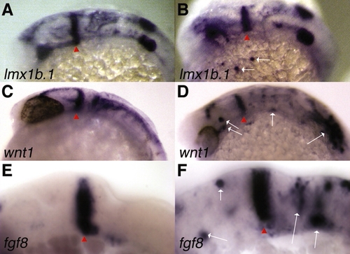
Heatshock-induced ectopic expression of lmx1b.1, wnt1 and fgf8. (B,D,F) One-cell stage embryos were injected with Hsp70-Lmx1b.1 (B,D) or Hsp70-Lmx1b.2 (F), heat shocked during early somitogenesis, and fixed approximately 12 hours after heat shock. Embryos were then examined for lmx1b.1, wnt1 or fgf8 expression by whole-mount in situ hybridization. (A,C,E) Uninjected, heat-shocked embryos were used as controls. Lateral views with anterior leftwards and dorsal upwards are shown. Red arrowheads indicate position of MMR and white arrows indicate ectopic clones.
|

Unillustrated author statements EXPRESSION / LABELING:
|