
|
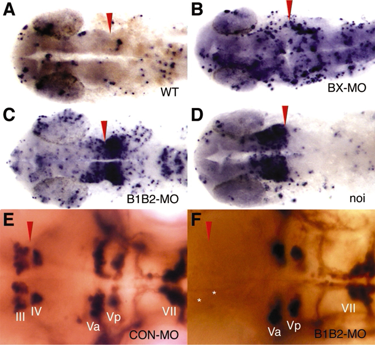
Fig. 7 Knockdown of Lmx1b.1 and Lmx1b.2 results in increased cell death around the IsO, with a concomitant failure of neural differentiation. (A-D) A TUNEL assay stains apoptotic cells blue. Dorsal views with anterior leftwards. (A) A low level of randomly distributed apoptosis is detected in wild-type embryos. (B) Injection of BX-MO results in a nonspecific increase in apoptosis. (C) Injection of B1B2-MO sharply increases apoptosis in the IsO, especially caudally. Nonspecific apoptosis is also increased. (D) Increased apoptosis is detected around the IsO in embryos homozygous for the pax2.1/no isthmus mutation, especially rostrally. (E,F) Cranial motoneurons were visualized by expressing GFP under the control of the Islet1 promoter and staining with an antibody against GFP. (E) Cranial motoneurons III and IV flank the IsO. (F) Neurons III and IV fail to form in B1B2-MO-injected embryos, while other neurons are unaffected. Red arrowheads indicate position of the MMR and asterisks mark normal position of missing neurons.