
Fig. 2
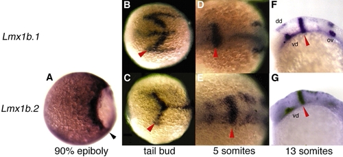
Expression of lmx1b.1 and lmx1b.2 in wild-type embryos. (A) Lateral view of a gastrula at 90% epiboly, animal pole leftwards. lmx1b.2 is expressed in the blastoderm margin (black arrowhead). (B-E) Dorsal views with anterior towards the left. (B,C) At the tailbud stage, lmx1b.1 and lmx1b.2 are both expressed in chevron-shaped domains at the presumptive MMR. Both are also expressed in the midline, with lmx1b.1 extending more rostrally and lmx1b.2 extending more caudally. (D,E) By the five-somite stage, expression of lmx1b.1 and lmx1b.2 are refined to rings at the MMR. lmx1b.1 is now expressed at the developing otic placode, while lmx1b.2 is expressed in the diencephalic region and in cells of the hindbrain that are converging on the dorsal midline. (F,G) Lateral views, rostral leftwards. By the 13-somite stage, lmx1b.1 and lmx1b.2 are both stably expressed at the MMR, the ventral diencephalon and the dorsal midline of the caudal CNS. lmx1b.1 is also stably expressed in the otic placodes. Red arrowheads indicate the MMR. ov, otic vesicle; dd, dorsal diencephalon; vd, ventral diencephalon.
|
| Genes: | |
|---|---|
| Fish: | |
| Knockdown Reagents: | |
| Anatomical Terms: | |
| Stage Range: | 90%-epiboly to 14-19 somites |