- Title
-
The zebrafish bozozok locus encodes Dharma, a homeodomain protein essential for induction of gastrula organizer and dorsoanterior embryonic structures
- Authors
- Fekany, K., Yamanaka, Y., Leung, T., Sirotkin, H.I., Topczewski, J., Gates, M.A., Hibi, M., Renucci, A., Stemple, D., Radbill, A., Schier, A.F., Driever, W., Hirano, T., Talbot, W.S., and Solnica-Krezel, L.
- Source
- Full text @ Development
|
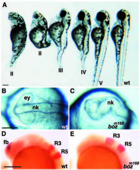
bozm168 causes deficiencies in dorsal and anterior embryonic structures with variable penetrance and expressivity. (A) boz mutant classes. From right to left, lateral views of wild-type, class V, class IV, class III and two class II boz larvae (3 dpf). There is a break in the notochord of class V boz larva, while the class IV mutant has a gap in notochord, spanning many somites. The class III mutant exhibits a gap in notochord and partially fused eyes, while class II mutants lack notochord completely and display cyclopia. (B,C) The neural keel in class II boz mutant embryos is highly abnormal, and the most anterior structures are missing (C), compared to wild type (B) (10-somite stage). (D,E) In wild-type embryos, emx1 (blue) is expressed in the forebrain (fb) and krox20 (red) in hindbrain rhombomeres R3 and R5 (D). In bozm168 class I embryos, emx1 expression is absent, while the krox20 expression domains appear enlarged (E) (17 hpf, lateral views). Scale bar, 200 μm. EXPRESSION / LABELING:
PHENOTYPE:
|
|
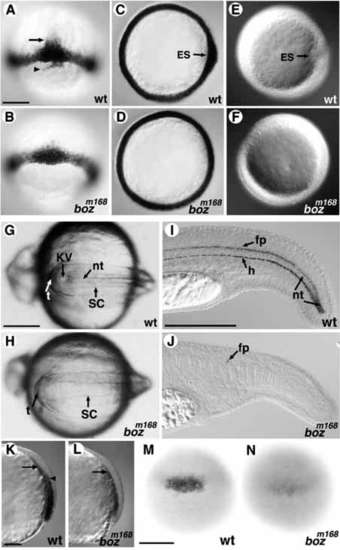
Development of axial tissues is affected by the bozm168 mutation. (A-D) (60% epiboly) ntl expression around the blastoderm margin is similar in wild-type (A) and boz embryos (B). ntl expression in notochord precursors above the margin (arrow), and in dorsal forerunner cells below the margin (arrowhead) in wild type (A) is reduced/absent in boz (B). The thickening of ntl expression seen in the animal view on the dorsal side of wild-type embryos (C) is not observed in bozm168 mutants (D). (E,F) The embryonic shield (ES) forms as a dorsal thickening of the germ ring in wild-type embryos (E), but it does not form in boz mutants (F). (G,H) During somitogenesis, Kupffer’s vesicle is seen posterior to the notochord in wild-type embryos at 10 somites. (H) Both Kupffer’s vesicle and the notochord are absent in bozm168 mutant siblings. (I,J) col2a1 expression in floor plate, hypochord, and nascent notochord at 1 dpf in the wild-type tail. All of these expression domains are absent, with exception of a few floor plate cells, in bozm168 mutants (J). (K,L) axial expression at 70% epiboly is normally seen in the endoderm (closest to the yolk, arrow) and dorsal mesoderm (arrowhead in K). In bozm168 mutant gastrulae, mesodermal but not endodermal axial expression is reduced/absent (arrow in L). Lateral views with dorsal to the right. (M,N) Dorsal flh expression in wildtype embryos (M) is decreased in bozm168 embryos at the germ ring stage (N). ES, embryonic shield; KV, Kupffer’s vesicle; nt, notochord; sc, spinal cord; t, tailbud; fp, floor plate; h, hypochord. Scale bar, 200 μm. PHENOTYPE:
|
|
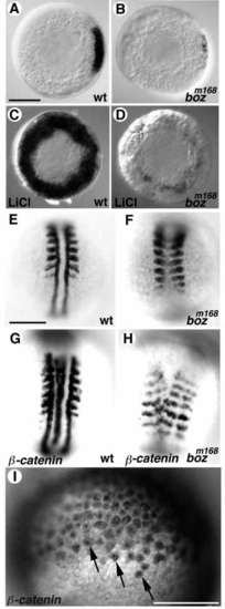
boz acts downstream or in parallel to the maternal β-catenin pathway. (A,B) gsc expression in the dorsal margin of wild-type embryos (A) is reduced in boz mutants (B). (C,D) After treatment of embryos with LiCl, gsc is ectopically expressed at high levels around the blastoderm margin in wild-type (C), but only at very low levels in putative boz mutant siblings (D). (E,F) myoD expression in untreated wild-type embryos is detected in the somites and adaxial cells (E). In bozm168 mutants, somitic myoD expression is fused and adaxial expression is absent (F). (G,H) After overexpression of b-catenin, wild-type embryos exhibit two complete arrays of myoD expression (G), while boz mutants have incomplete duplicated arrays of myoD expression with fused somites and absent adaxial cells (H). (I) Distribution of β-catenin at the blastula stage after injection of b- catenin mRNA. β-catenin (signal is brown) is detected in the cytoplasm and at higher levels in nuclei of numerous blastomeres (arrows). (A-D) Animal views; (A,B) Dorsal to the right; (E-I) Dorsal view, anterior to the top. Scale bar, 200 μm. EXPRESSION / LABELING:
|
|
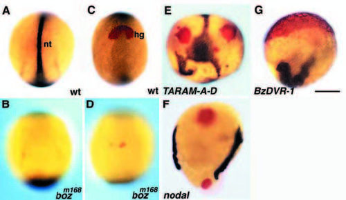
TGF-β signaling suppresses the boz phenotype. (A,B) ntl expression (blue) normally marks notochord precursors (nt) and tail bud in wild-type embryos (A). In boz embryos, ntl expression in notochord precursors is absent (B) (5-7 somites). (C,D hgg1 expression (red) in the prospective hatching gland (hg) of a wild-type embryo (C) is reduced in bozm168 mutants (D). (EG) Overexpression of TARAM-A-D (E), murine Nodal (F) and BMP-Vg1 (G) resulted in ectopic ntl and hgg1 expression domains in the majority of embryos from wild-type and bozm168/m168 parents. (A,B) Dorsal view, (C,D) Frontal view; in all images anterior is to the top. Scale bar, 200 μm. EXPRESSION / LABELING:
|
|
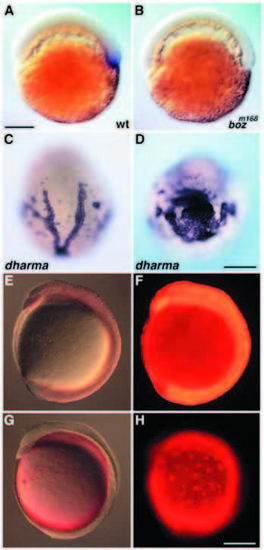
Analysis of expression and rescuing potential of dharma RNA in boz mutants. (A,B) Whole-mount in situ hybridization reveals expression of dharma in the dorsal YSL of wild-type embryos at 50% epiboly (A). dharma was not expressed in sibling boz embryos (B). (C,D) Expanded ntl and hgg1 expression in embryos from bozm168/+ parents injected with dharma at the 1- to 4-cell stage. (E,F) Embryos coinjected with dharma and fluorescent dextran into one cell at the 16-cell stage. Nomarski (E) and fluorescent (F) images reveal that dextran is localized to the blastoderm. (G,H) After injections into the yolk at 1000-cell stage, fluorescent dextran remains confined to the YSL as seen in a Nomarski (G) and fluorescent (H) image. (A,B,EH) Lateral views with animal pole to the top, (E-H) dorsal to the right; (C) animal view. Scale bar, 200 μm. EXPRESSION / LABELING:
|

