
|
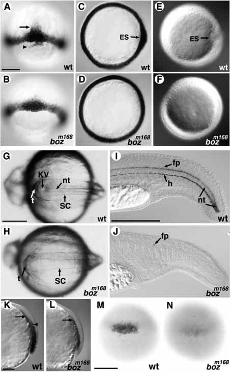
Fig. 2 Development of axial tissues is affected by the bozm168 mutation. (A-D) (60% epiboly) ntl expression around the blastoderm margin is similar in wild-type (A) and boz embryos (B). ntl expression in notochord precursors above the margin (arrow), and in dorsal forerunner cells below the margin (arrowhead) in wild type (A) is reduced/absent in boz (B). The thickening of ntl expression seen in the animal view on the dorsal side of wild-type embryos (C) is not observed in bozm168 mutants (D). (E,F) The embryonic shield (ES) forms as a dorsal thickening of the germ ring in wild-type embryos (E), but it does not form in boz mutants (F). (G,H) During somitogenesis, Kupffer’s vesicle is seen posterior to the notochord in wild-type embryos at 10 somites. (H) Both Kupffer’s vesicle and the notochord are absent in bozm168 mutant siblings. (I,J) col2a1 expression in floor plate, hypochord, and nascent notochord at 1 dpf in the wild-type tail. All of these expression domains are absent, with exception of a few floor plate cells, in bozm168 mutants (J). (K,L) axial expression at 70% epiboly is normally seen in the endoderm (closest to the yolk, arrow) and dorsal mesoderm (arrowhead in K). In bozm168 mutant gastrulae, mesodermal but not endodermal axial expression is reduced/absent (arrow in L). Lateral views with dorsal to the right. (M,N) Dorsal flh expression in wildtype embryos (M) is decreased in bozm168 embryos at the germ ring stage (N). ES, embryonic shield; KV, Kupffer’s vesicle; nt, notochord; sc, spinal cord; t, tailbud; fp, floor plate; h, hypochord. Scale bar, 200 μm.