
|
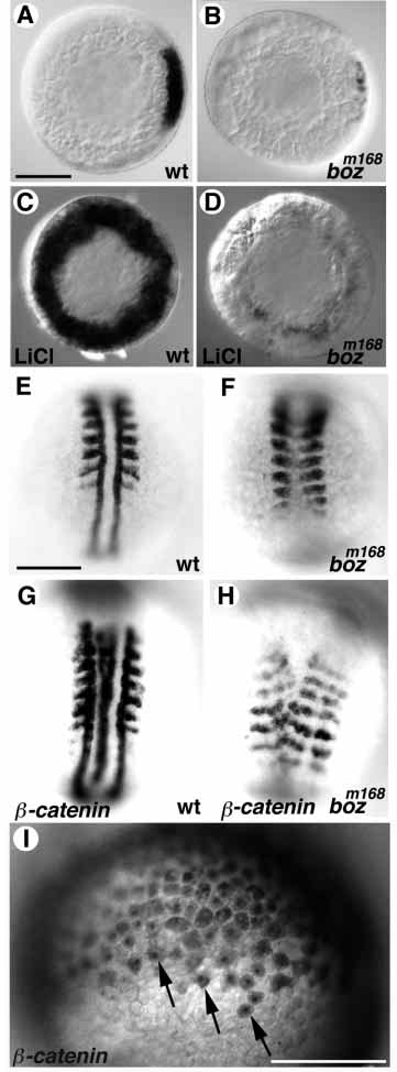
Fig. 3 boz acts downstream or in parallel to the maternal β-catenin pathway. (A,B) gsc expression in the dorsal margin of wild-type embryos (A) is reduced in boz mutants (B). (C,D) After treatment of embryos with LiCl, gsc is ectopically expressed at high levels around the blastoderm margin in wild-type (C), but only at very low levels in putative boz mutant siblings (D). (E,F) myoD expression in untreated wild-type embryos is detected in the somites and adaxial cells (E). In bozm168 mutants, somitic myoD expression is fused and adaxial expression is absent (F). (G,H) After overexpression of b-catenin, wild-type embryos exhibit two complete arrays of myoD expression (G), while boz mutants have incomplete duplicated arrays of myoD expression with fused somites and absent adaxial cells (H). (I) Distribution of β-catenin at the blastula stage after injection of b- catenin mRNA. β-catenin (signal is brown) is detected in the cytoplasm and at higher levels in nuclei of numerous blastomeres (arrows). (A-D) Animal views; (A,B) Dorsal to the right; (E-I) Dorsal view, anterior to the top. Scale bar, 200 μm.