- Title
-
Mutations affecting the cardiovascular system and other internal organs in zebrafish
- Authors
- Chen, J.N., Haffter, P., Odenthal, J., Vogelsang, E., Brand, M., van Eeden, F.J., Furutani-Seiki, M., Granato, M., Hammerschmidt, M., Heisenberg, C.P., Jiang, Y.J., Kane, D.A., Kelsh, R.N., Mullins, M.C., and Nüsslein-Volhard, C.
- Source
- Full text @ Development
|
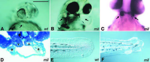
Cardia bifida mutants. Two hearts are located at either side of the embryo. A ventral view of 1-day old embryos shows two swollen pericardial sac in milte273 (B) but not in the wild-type sibling (A). Two hearts can be clearly visualized either by immunostaining or in histological sections. (C) A 2-day old milte273 embryo stained with the antibody S46, which labels the atria. (D) A transverse section of a 2-day old milte273 embryo. The hearts are indicated by arrows; a, atrium; v, ventricle. Images of tails of live 1-day old wild-type and milte273 embryos are shown in E and F, respectively. Scale bar, 100 μm. |
|
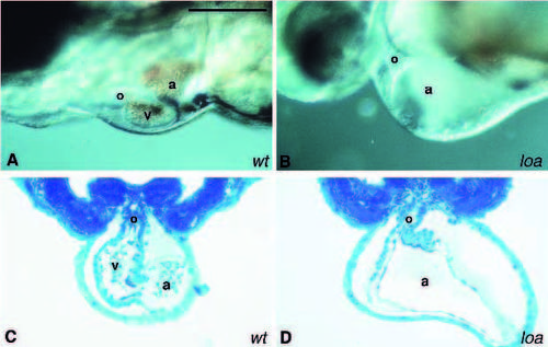
Chamber demarcation mutant. Images of the hearts of live 2-day old wild-type (A) and loatu29d (B) embryos are shown. The atrium (a) is significantly enlarged in loatu29d. No obvious structure representing the ventricle (v) is seen between the atrium and the outflow tract (o). The atrium, ventricle and outflow tract are present in a transverse section of the heart in a 2-day old wild-type embryo (C). However, no obvious structure representing the ventricle is seen in a transverse section of the heart of the 2-day old loatu29d embryo (D). The embryos are oriented with the anterior to the left and dorsal to the top. Scale bar, 100 μm. PHENOTYPE:
|
|
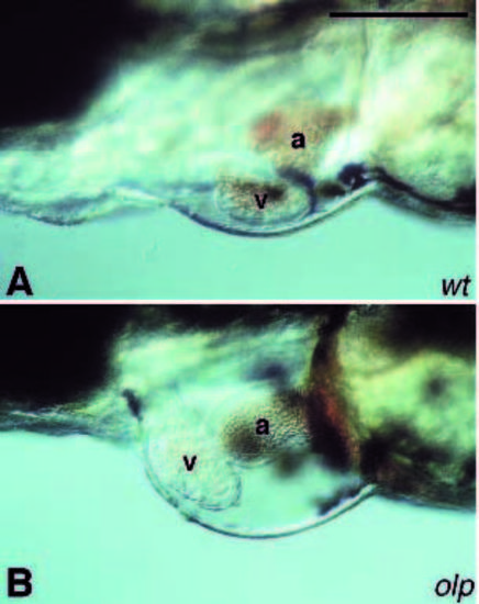
Chamber positioning mutant. Images of the heart of live 2-day old wildtype (A) and olp (B) embryos. The ventricle (v) is placed on top of the atrium (a) at a 90 degree angle in the olp mutant (B), whereas they are parallel to each other with the atrium on the left and the ventricle on the right side in the wildtype embryo (A). Scale bar, 100 μm. PHENOTYPE:
|
|
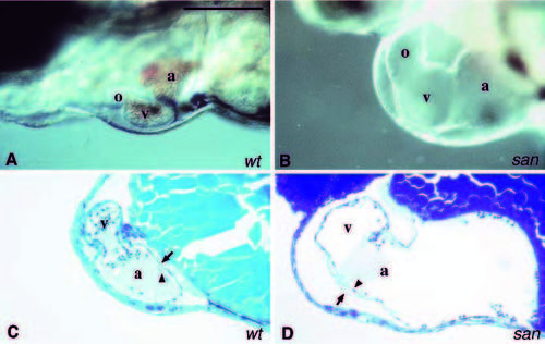
Mutant with an enlarged heart. Images of the hearts of live embryos. (A) Wild-type embryo. As shown, the atrium (a), ventricle (v) and the outflow tract (o) are located on the ventral side of the embryo. Looping places the atrium to the left side of the embryo. The circulation is strong and the heart is full of blood cells. (B) The heart of a 2-day old santy219c embryo. The atrium (a), ventricle (v) and the outflow tract (o) are significantly enlarged in this mutant. The absence of circulation results in a lack of blood cells in the heart chambers. Sagittal sections of the hearts of normal 2-day old wild-type (C) and santy219c (D) embryos. Because of the looping the chambers are placed in different focal planes so only the ventricle and the atrium are shown in these sections. The myocardium (arrow) and endocardium (arrowhead) are clearly distinguishable. The space between the myocardial and endocardial cell layers in the mutant atrium is much thinner than in the normal heart. Scale bar, 100 μm. PHENOTYPE:
|
|
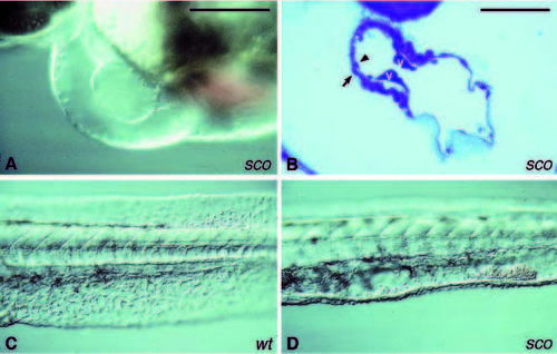
Mutants with reduced cardiac jelly. (A) Image of the heart of a live 2-day old scote382 embryo. Only the myocardial layer is visible in the atrium but not the endocardial layer. The low circulation results in a lack of blood cells in the heart. (B) A sagittal section of the heart of a 2- day old scote382 embryo. Both, the myocardium (arrow) and endocardium (arrowhead) are present in the heart. However, the matrix between the two cell layers is very thin. The endocardium seems to attach to the myocardium, especially in the atrium. Valves (v) are formed and placed at the atrial-ventricular junction. In addition, the endothelial cells in the ventral fin of 2-day old scote382 embryos (D) are not as organized as in the wild type siblings (C), and blood cells accumulate in the empty space in the ventral tail fin. Scale bar, (A,C and D) 100 μm; (B) 50 μm. |
|
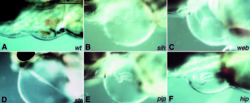
Abnormal heart morphology as a result of heart failure. The shape of the heart in the mutant embryos is often distorted as the result of the failing function. Images of the hearts of live 2-day old wild-type (A), sihte300b (B), webtc288b (C), strtu255b (D), piptq286e (E) and hiptx218 (F) embryos. Scale bar, 100 μm. PHENOTYPE:
|
|
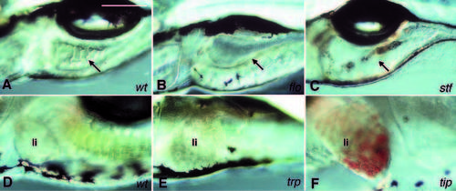
Intestine and liver mutants. (A-C) Images of the intestines of live 5-day old embryos. A thick epithelial cell layer with folds is the characteristic feature of the intestine in wild-type (A). Mutants of floti262c and stftz259 have a thin epithelial cell layer and lack the characteristic folds of the intestine (B,C). In addition, there is grey material in the gut of 5-day old stftz259 embryos (C). The arrows point to the intestine. (D-F) Images of the livers (li) of live 5-day old embryos. Mutants in the necrosing liver class have grey granules in the liver. The liver of trptm117d. embryos is shown in E. In tipth203 embryos, the liver accumulates red blood cells (F). Scale bar, 100 μm. PHENOTYPE:
|

ZFIN is incorporating published figure images and captions as part of an ongoing project. Figures from some publications have not yet been curated, or are not available for display because of copyright restrictions. PHENOTYPE:
|

Unillustrated author statements |

