
|
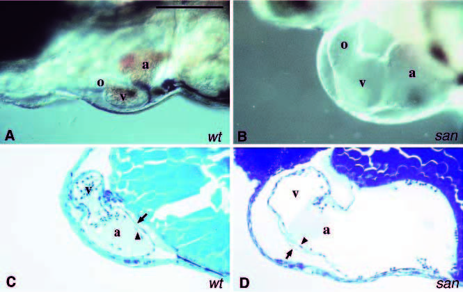
Fig. 4 Mutant with an enlarged heart. Images of the hearts of live embryos. (A) Wild-type embryo. As shown, the atrium (a), ventricle (v) and the outflow tract (o) are located on the ventral side of the embryo. Looping places the atrium to the left side of the embryo. The circulation is strong and the heart is full of blood cells. (B) The heart of a 2-day old santy219c embryo. The atrium (a), ventricle (v) and the outflow tract (o) are significantly enlarged in this mutant. The absence of circulation results in a lack of blood cells in the heart chambers. Sagittal sections of the hearts of normal 2-day old wild-type (C) and santy219c (D) embryos. Because of the looping the chambers are placed in different focal planes so only the ventricle and the atrium are shown in these sections. The myocardium (arrow) and endocardium (arrowhead) are clearly distinguishable. The space between the myocardial and endocardial cell layers in the mutant atrium is much thinner than in the normal heart. Scale bar, 100 μm.