
Fig. 5
- ID
- ZDB-IMAGE-140304-71
- Publication
- Chen et al., 1996 - Mutations affecting the cardiovascular system and other internal organs in zebrafish
- All Figures
- Figures for Chen et al., 1996
|
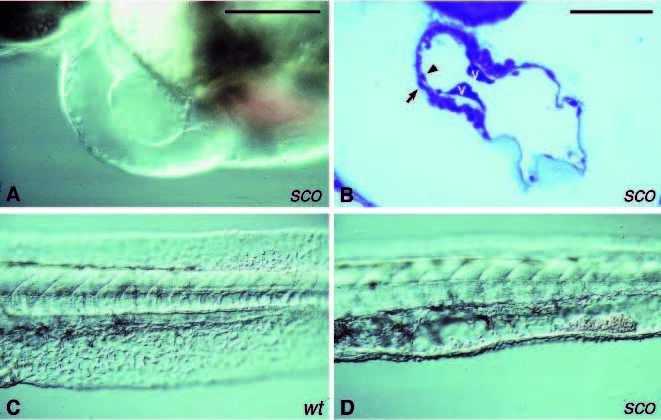
Fig. 5 Mutants with reduced cardiac jelly. (A) Image of the heart of a live 2-day old scote382 embryo. Only the myocardial layer is visible in the atrium but not the endocardial layer. The low circulation results in a lack of blood cells in the heart. (B) A sagittal section of the heart of a 2- day old scote382 embryo. Both, the myocardium (arrow) and endocardium (arrowhead) are present in the heart. However, the matrix between the two cell layers is very thin. The endocardium seems to attach to the myocardium, especially in the atrium. Valves (v) are formed and placed at the atrial-ventricular junction. In addition, the endothelial cells in the ventral fin of 2-day old scote382 embryos (D) are not as organized as in the wild type siblings (C), and blood cells accumulate in the empty space in the ventral tail fin. Scale bar, (A,C and D) 100 μm; (B) 50 μm.