FIGURE

Fig. 1

ID
ZDB-FIG-091202-2
Publication
Chen et al., 1996 - Mutations affecting the cardiovascular system and other internal organs in zebrafish
Other Figures
All Figure Page
Back to All Figure Page
Fig. 1

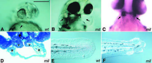
Cardia bifida mutants. Two hearts are located at either side of the embryo. A ventral view of 1-day old embryos shows two swollen pericardial sac in milte273 (B) but not in the wild-type sibling (A). Two hearts can be clearly visualized either by immunostaining or in histological sections. (C) A 2-day old milte273 embryo stained with the antibody S46, which labels the atria. (D) A transverse section of a 2-day old milte273 embryo. The hearts are indicated by arrows; a, atrium; v, ventricle. Images of tails of live 1-day old wild-type and milte273 embryos are shown in E and F, respectively. Scale bar, 100 μm.

Expression Data
Antibody:
Fish:
Anatomical Term:
Stage: Long-pec

Expression Detail
Antibody Labeling
Phenotype Data
Fish:
Observed In:
Stage Range: Prim-5 to Long-pec

Phenotype Detail
Acknowledgments
This image is the copyrighted work of the attributed author or publisher, and ZFIN has permission only to display this image to its users. Additional permissions should be obtained from the applicable author or publisher of the image. Full text @ Development