
Fig. 7
- ID
- ZDB-IMAGE-140304-74
- Publication
- Chen et al., 1996 - Mutations affecting the cardiovascular system and other internal organs in zebrafish
- All Figures
- Figures for Chen et al., 1996
|
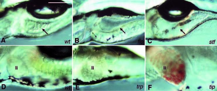
Fig. 7 Intestine and liver mutants. (A-C) Images of the intestines of live 5-day old embryos. A thick epithelial cell layer with folds is the characteristic feature of the intestine in wild-type (A). Mutants of floti262c and stftz259 have a thin epithelial cell layer and lack the characteristic folds of the intestine (B,C). In addition, there is grey material in the gut of 5-day old stftz259 embryos (C). The arrows point to the intestine. (D-F) Images of the livers (li) of live 5-day old embryos. Mutants in the necrosing liver class have grey granules in the liver. The liver of trptm117d. embryos is shown in E. In tipth203 embryos, the liver accumulates red blood cells (F). Scale bar, 100 μm.