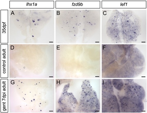
Expression of Wnt signaling pathway genes during zebrafish mesonephric kidney development and adult regeneration. (A-C) Whole-mount in situ hybridization showing the trunk kidney region of juvenile zebrafish at 35 days post fertilization (dpf). Juvenile zebrafish are rapidly growing and adding new nephrons. The transcription factor lhx1a is expressed in cell aggregates comprising newly forming nephrons (A), while the canonical Wnt receptor fzd9b (B) and canonical Wnt transcription factor lef1 (C) appear in similar aggregates, as well as in smaller clusters and single cells. (D-I) Whole-mount in situ hybridization showing the anterior trunk kidney region of adult zebrafish (between 6 months and 2 years). (D-F) Adult zebrafish are no longer actively forming new nephrons and only rarely express lhx1a, fzd9b or lef1 in aggregates. (G-I) In response to acute kidney injury by gentamicin injection, nephron formation is reinitiated by 7 days post-injury (dpi), and lhx1a, fzd9b and lef1 are strongly expressed in large aggregates and small clusters of cells. Representative images from n=12 (lhx1a), n=9 (fzd9b) and n=10 (lef1) juveniles, and n=3 (uninj lhx1a), n=13 (7 dpi lhx1a), n=3 (uninj fzd9b), n=5 (7 dpi fzd9b), n=3 (uninj lef1) and n=5 (7 dpi lef1) adult fish per condition from two independent experiments. Scale bars: 0.1 mm in A-C; 0.2 mm in D-I.
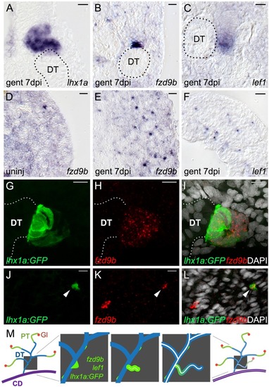
fzd9b and lef1 are expressed in regions overlapping the known nephron progenitor lhx1a. (A-F) High-magnification DIC images of sections from whole-mount in situ hybridized kidneys at the indicated time points. (A) The known nephron progenitor marker lhx1a is expressed in a new nephron extending from the junction with a pre-existing distal tubule (DT, outline). (B) fzd9b is restricted to aggregates and rosettes adjacent to existing distal tubules (DT, outline), while (C) lef1 extends along the distal end of new nephrons similarly to lhx1a. (D) fzd9b is expressed in rare single mesenchymal cells in the cortical interstitium of uninjured adult zebrafish kidneys and this population is increased after injury (E). (F) lef1 is also expressed in many single interstitial cells after injury. (G-L) Confocal images of immunostained whole-mount kidneys from the Tg(lhx1a:GFP) reporter line in which nephron progenitors are marked by GFP expression. Fluorescent in situ hybridization for fzd9b in red and cell nuclei are visualized with DAPI in white. (G-I) 7 days after gentamicin injury, lhx1a+ aggregates marked by GFP also express fzd9b. Confocal projection showing a rosette structure in contact with the existing distal tubule (white dotted line). (J-L) Confocal slice showing that single mesenchymal lhx1a+ cells (arrowheads) also express fzd9b. (M) Diagram showing stages of new nephron formation from adult kidney stem cells. The adult mesonephric kidney structure showing glomeruli (Gl; red), proximal tubule (PT; green), distal tubule (DT; blue) and collecting duct (CD; purple). Enlarged view of distal tubules (blue) shows sequential steps of new nephron formation initiated with the formation of a polarized rosette of aggregated renal stem cells (green), outgrowth of progenitors to form a primitive tubule, lumen fusion and differentiation of the new nephron (leading to nephron segmentation), and finally development of a filtering fully functional nephron (Diep et al., 2011). Scale bars: 10 µm.
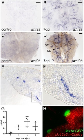
wnt9a and wnt9b are induced in distal tubules and collecting ducts after injury. (A,B) Whole-mount in situ hybridization for wnt9a. (A) wnt9a is expressed at very low levels in uninjured adult kidney tubular epithelium. (B) By 7 dpi, wnt9a is strongly induced in the branched distal tubule segments of nephrons. (C) wnt9b is expressed at low levels in uninjured adult kidney tubular epithelium. (D) After 7 dpi, wnt9b is strongly induced in the branched distal tubule segments of nephrons (DT; arrowheads) and common collecting ducts (CDs; arrow). (E) DIC image of a section through a 7 dpi kidney showing wnt9b expression in cross-sections of tubular epithelium. (F) Higher-magnification image of boxed area shown in E with wnt9b expression in a lengthwise section of tubule. (G) Quantification by qPCR at the indicated time points after injury shows that wnt9b expression was increased by 3 dpi and peaked at 5 dpi. Data derived from three individual fish per time point as indicated by individual graph symbols. Data are mean±s.d. (H) Confocal stack projection of Tg(lhx1a:GFP)×Tg(slc12a3:mCherry) transgenic kidney tissue. lhx1a:GFP nephron aggregates form exclusively on slc12a3:mCherry-positive distal tubules. Representative images from n=2 (A), n=3 (B), n=6 (C), n=4 (D), n=3 [H, Tg(lhx1a:GFP)×Tg(slc12a3:mCherry)] fish per condition. Scale bars: 0.2 mm in A-F; 10 µm in H.
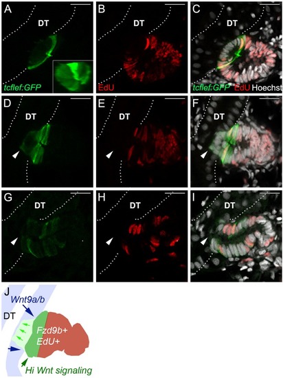
New nephron aggregates are patterned by canonical Wnt activity that defines a zone of cell proliferation. (A) Tg(TCF/Lef-miniP:dGFP) reporter expression (green) is restricted to a single-cell thick dome of cells, reflecting a domain of high canonical Wnt activity in new nephron aggregates. (B) EdU incorporation (red) in nephron aggregates. (C) Merged image shows cells adjacent to existing tubules are not proliferating, whereas cells in the Wnthigh dome and at a distance from existing tubules are highly proliferative. (D-F) An example of a new nephron that has invaded the distal tubule (arrowheads). Invading cells are Wntlow, EdU−, whereas Wnthigh, EdU+ cells appear flush with the basal surface of the existing distal tubule. (G-I) A new nephron with lumenal connection to the existing distal tubule (arrowheads). Regionalized expression of GFP has been lost and the number of EdU+ nuclei has decreased. (J) Diagram of a nephron aggregate at the stage of tubule invasion (F) labeled to show sources of Wnt9a and Wntb (distal tubule, DT; blue, blue arrows), invading cells (green arrows), high canonical Wnt signaling cells (green) and the localization of Frizzled 9b-positive new nephron cells that are also EdU-positive (brown). White dotted lines outline the existing distal tubules (DT). Hoechst labels nuclei. Representative images from n=6 fish. Scale bars: 10 µm.
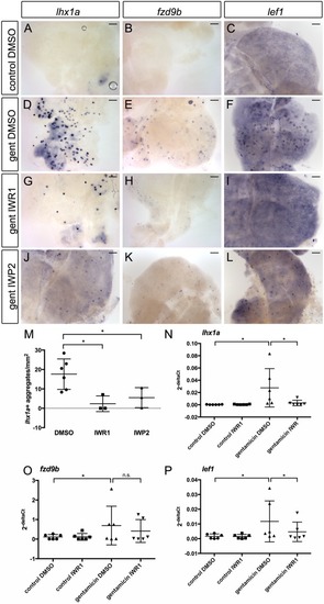
Wnt inhibition blocks new nephron formation. Gentamicin-injured adult zebrafish were treated with either DMSO or 5 µM of the Wnt inhibitors IWR1 or IWP2 in system water starting at 1 dpi. Whole-mount in situ hybridization showing the trunk kidney region at 7 dpi. (A-C) Control-injected and DMSO-treated kidneys do not express markers of new nephrons. (D-F) Injury induces cell aggregates expressing lhx1a, fzd9b and lef1. Nephron aggregate formation is blocked by Wnt inhibition using IWR1 (G-I) or IWP2 (J-L). Scale bars: 0.2 mm. (M-P) Quantification of Wnt inhibitor effects on lhx1a+ aggregates and gene expression. (M) Percentage of lhx1a+ aggregates/mm2 of kidney calculated using ImageJ. n=3-6 fish for each condition, as indicated by individual graph symbols. (N-P) qPCR quantification of lhx1a, fzd9b and lef1 mRNA in kidney tissue harvested 7 d after gentamicin injury, n=6 for each condition. *P<0.05 calculated using Student's unpaired two-tailed t-test. Data are mean±s.d.
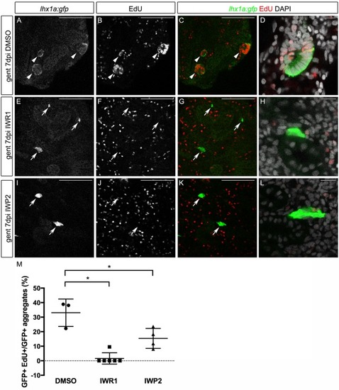
Wnt inhibition blocks proliferation in nephron aggregates.Tg(lhx1a:GFP) transgenic fish expressing GFP in aggregates and new nephrons were injured by gentamicin injection, injected with EdU to label proliferating nuclei at 6 dpi and kidneys were harvested at 7 dpi. Single slices from confocal z-stacks are shown. (A-D) Gentamicin induces GFP+ new nephrons with proliferating EdU+ nuclei. (E-L) Inhibition of Wnt signaling leads to a loss of proliferation and no morphological sign of nephron formation. GFP+ aggregates are still visible adjacent to existing tubules; however, organized structures, such as rosettes or polarized proliferating new nephrons, were rarely observed. Arrows indicate double-labeled new nephrons. Scale bars: 100 µm in A-C,E-G,I-K; 10 µm in D,H,L. (M) Quantification of GFP+ aggregates with more than five EdU+ nuclei expressed as a percentage of total GFP+ aggregates. n=3-6 fish for each condition, as indicated by graph symbols. n=4-7 confocal z-stacks from each kidney. *P<0.05 calculated using Student's unpaired two-tailed t-test. Data are mean±s.d.
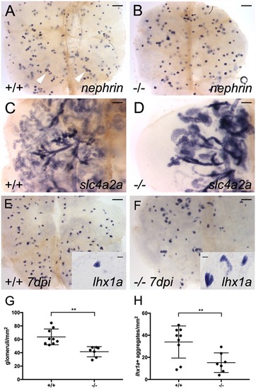
Mutation in fzd9b reduces nephron number and inhibits nephron regeneration. (A) Whole-mount in situ hybridization with a nephrin antisense mRNA probe reveals nephron glomeruli in wild-type adult kidney tissue. Clusters of up to six glomeruli are visible (white arrowheads). (B) In maternal zygotic (MZ) fzd9bfb203 homozygous mutants raised to adulthood, nephrin in situ hybridization reveals fewer widely scattered glomeruli in adult kidney tissue (quantified in G). (C) slc4a2a in situ hybridization reveals the branched kidney nephron structure in the wild-type adult kidney. (D) In adult MZ fzd9bfb203 homozygous mutants, kidney tissue shows a marked reduction in the number of nephron tubules revealed by slc4a2a in situ expression [all fzd9b−/− mutants showed a decrease in tubules (n=4) compared with unrelated wild type (n=3)]. (E) Seven day post-gentamicin injury kidney shows robust production of lhx1a-positive new nephron aggregates. (F) MZ fzd9bfb203−/− adult kidney 7 days post-injury shows markedly reduced lhx1a-positive new nephron aggregates. (G) Quantification of nephrin-positive glomeruli per mm2 shows a roughly 30% reduction in fzd9bfb203−/− mutant kidneys. Data represent results from seven to nine individual fish, as indicated by graph symbols. (H) Quantification of lhx1a-positive new nephron aggregates 7 days post-injury shows a marked reduction in fzd9bfb203−/− adult kidney. Student's unpaired two-tailed t-test, **P<0.01. Data are mean±s.d. Scale bars: 0.2 mm in A,B,E,F; 0.1 mm in C,D; 10 µm in insets in E,F.
Acknowledgments
This image is the copyrighted work of the attributed author or publisher, and
ZFIN has permission only to display this image to its users.
Additional permissions should be obtained from the applicable author or publisher of the image.
Full text @ Development
Your Input Welcome
Thank you for submitting comments. Your input has been emailed to ZFIN curators who may contact you if
additional information is required.
Oops. Something went wrong. Please try again later.