IMAGE

Fig. 7.

ID
ZDB-IMAGE-190723-1999
Source
Figures for Kamei et al., 2019
Image
Figure Caption

Fig. 7.

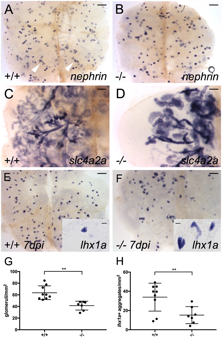
Mutation in fzd9b reduces nephron number and inhibits nephron regeneration. (A) Whole-mount in situ hybridization with a nephrin antisense mRNA probe reveals nephron glomeruli in wild-type adult kidney tissue. Clusters of up to six glomeruli are visible (white arrowheads). (B) In maternal zygotic (MZ) fzd9bfb203 homozygous mutants raised to adulthood, nephrin in situ hybridization reveals fewer widely scattered glomeruli in adult kidney tissue (quantified in G). (C) slc4a2a in situ hybridization reveals the branched kidney nephron structure in the wild-type adult kidney. (D) In adult MZ fzd9bfb203 homozygous mutants, kidney tissue shows a marked reduction in the number of nephron tubules revealed by slc4a2a in situ expression [all fzd9b−/− mutants showed a decrease in tubules (n=4) compared with unrelated wild type (n=3)]. (E) Seven day post-gentamicin injury kidney shows robust production of lhx1a-positive new nephron aggregates. (F) MZ fzd9bfb203−/− adult kidney 7 days post-injury shows markedly reduced lhx1a-positive new nephron aggregates. (G) Quantification of nephrin-positive glomeruli per mm2 shows a roughly 30% reduction in fzd9bfb203−/− mutant kidneys. Data represent results from seven to nine individual fish, as indicated by graph symbols. (H) Quantification of lhx1a-positive new nephron aggregates 7 days post-injury shows a marked reduction in fzd9bfb203−/− adult kidney. Student's unpaired two-tailed t-test, **P<0.01. Data are mean±s.d. Scale bars: 0.2 mm in A,B,E,F; 0.1 mm in C,D; 10 µm in insets in E,F.

Acknowledgments
This image is the copyrighted work of the attributed author or publisher, and ZFIN has permission only to display this image to its users. Additional permissions should be obtained from the applicable author or publisher of the image. Full text @ Development