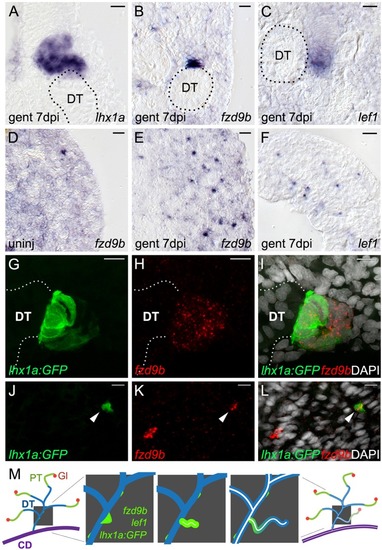
fzd9b and lef1 are expressed in regions overlapping the known nephron progenitor lhx1a. (A-F) High-magnification DIC images of sections from whole-mount in situ hybridized kidneys at the indicated time points. (A) The known nephron progenitor marker lhx1a is expressed in a new nephron extending from the junction with a pre-existing distal tubule (DT, outline). (B) fzd9b is restricted to aggregates and rosettes adjacent to existing distal tubules (DT, outline), while (C) lef1 extends along the distal end of new nephrons similarly to lhx1a. (D) fzd9b is expressed in rare single mesenchymal cells in the cortical interstitium of uninjured adult zebrafish kidneys and this population is increased after injury (E). (F) lef1 is also expressed in many single interstitial cells after injury. (G-L) Confocal images of immunostained whole-mount kidneys from the Tg(lhx1a:GFP) reporter line in which nephron progenitors are marked by GFP expression. Fluorescent in situ hybridization for fzd9b in red and cell nuclei are visualized with DAPI in white. (G-I) 7 days after gentamicin injury, lhx1a+ aggregates marked by GFP also express fzd9b. Confocal projection showing a rosette structure in contact with the existing distal tubule (white dotted line). (J-L) Confocal slice showing that single mesenchymal lhx1a+ cells (arrowheads) also express fzd9b. (M) Diagram showing stages of new nephron formation from adult kidney stem cells. The adult mesonephric kidney structure showing glomeruli (Gl; red), proximal tubule (PT; green), distal tubule (DT; blue) and collecting duct (CD; purple). Enlarged view of distal tubules (blue) shows sequential steps of new nephron formation initiated with the formation of a polarized rosette of aggregated renal stem cells (green), outgrowth of progenitors to form a primitive tubule, lumen fusion and differentiation of the new nephron (leading to nephron segmentation), and finally development of a filtering fully functional nephron (Diep et al., 2011). Scale bars: 10 µm.
|
