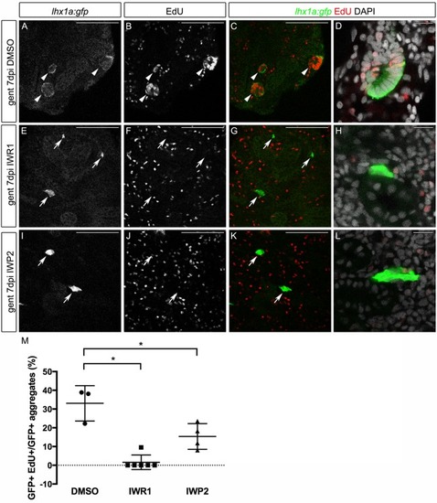
Wnt inhibition blocks proliferation in nephron aggregates.Tg(lhx1a:GFP) transgenic fish expressing GFP in aggregates and new nephrons were injured by gentamicin injection, injected with EdU to label proliferating nuclei at 6 dpi and kidneys were harvested at 7 dpi. Single slices from confocal z-stacks are shown. (A-D) Gentamicin induces GFP+ new nephrons with proliferating EdU+ nuclei. (E-L) Inhibition of Wnt signaling leads to a loss of proliferation and no morphological sign of nephron formation. GFP+ aggregates are still visible adjacent to existing tubules; however, organized structures, such as rosettes or polarized proliferating new nephrons, were rarely observed. Arrows indicate double-labeled new nephrons. Scale bars: 100 µm in A-C,E-G,I-K; 10 µm in D,H,L. (M) Quantification of GFP+ aggregates with more than five EdU+ nuclei expressed as a percentage of total GFP+ aggregates. n=3-6 fish for each condition, as indicated by graph symbols. n=4-7 confocal z-stacks from each kidney. *P<0.05 calculated using Student's unpaired two-tailed t-test. Data are mean±s.d.
|
