IMAGE

Fig. 3.

ID
ZDB-IMAGE-190723-1995
Source
Figures for Kamei et al., 2019
Image
Figure Caption

Fig. 3.

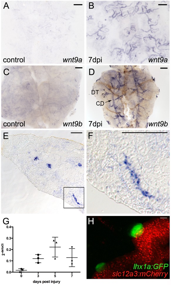
wnt9a and wnt9b are induced in distal tubules and collecting ducts after injury. (A,B) Whole-mount in situ hybridization for wnt9a. (A) wnt9a is expressed at very low levels in uninjured adult kidney tubular epithelium. (B) By 7 dpi, wnt9a is strongly induced in the branched distal tubule segments of nephrons. (C) wnt9b is expressed at low levels in uninjured adult kidney tubular epithelium. (D) After 7 dpi, wnt9b is strongly induced in the branched distal tubule segments of nephrons (DT; arrowheads) and common collecting ducts (CDs; arrow). (E) DIC image of a section through a 7 dpi kidney showing wnt9b expression in cross-sections of tubular epithelium. (F) Higher-magnification image of boxed area shown in E with wnt9b expression in a lengthwise section of tubule. (G) Quantification by qPCR at the indicated time points after injury shows that wnt9b expression was increased by 3 dpi and peaked at 5 dpi. Data derived from three individual fish per time point as indicated by individual graph symbols. Data are mean±s.d. (H) Confocal stack projection of Tg(lhx1a:GFP)×Tg(slc12a3:mCherry) transgenic kidney tissue. lhx1a:GFP nephron aggregates form exclusively on slc12a3:mCherry-positive distal tubules. Representative images from n=2 (A), n=3 (B), n=6 (C), n=4 (D), n=3 [H, Tg(lhx1a:GFP)×Tg(slc12a3:mCherry)] fish per condition. Scale bars: 0.2 mm in A-F; 10 µm in H.

Acknowledgments
This image is the copyrighted work of the attributed author or publisher, and ZFIN has permission only to display this image to its users. Additional permissions should be obtained from the applicable author or publisher of the image. Full text @ Development