IMAGE

Fig. 4.

ID
ZDB-IMAGE-190723-1996
Source
Figures for Kamei et al., 2019
Image
Figure Caption

Fig. 4.

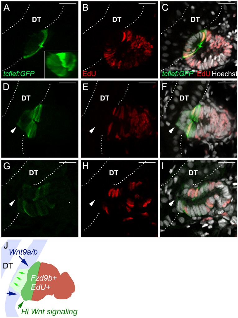
New nephron aggregates are patterned by canonical Wnt activity that defines a zone of cell proliferation. (A) Tg(TCF/Lef-miniP:dGFP) reporter expression (green) is restricted to a single-cell thick dome of cells, reflecting a domain of high canonical Wnt activity in new nephron aggregates. (B) EdU incorporation (red) in nephron aggregates. (C) Merged image shows cells adjacent to existing tubules are not proliferating, whereas cells in the Wnthigh dome and at a distance from existing tubules are highly proliferative. (D-F) An example of a new nephron that has invaded the distal tubule (arrowheads). Invading cells are Wntlow, EdU, whereas Wnthigh, EdU+ cells appear flush with the basal surface of the existing distal tubule. (G-I) A new nephron with lumenal connection to the existing distal tubule (arrowheads). Regionalized expression of GFP has been lost and the number of EdU+ nuclei has decreased. (J) Diagram of a nephron aggregate at the stage of tubule invasion (F) labeled to show sources of Wnt9a and Wntb (distal tubule, DT; blue, blue arrows), invading cells (green arrows), high canonical Wnt signaling cells (green) and the localization of Frizzled 9b-positive new nephron cells that are also EdU-positive (brown). White dotted lines outline the existing distal tubules (DT). Hoechst labels nuclei. Representative images from n=6 fish. Scale bars: 10 µm.

Acknowledgments
This image is the copyrighted work of the attributed author or publisher, and ZFIN has permission only to display this image to its users. Additional permissions should be obtained from the applicable author or publisher of the image. Full text @ Development