
- Title
-
Split top: A maternal cathepsin B that regulates dorsoventral patterning and morphogenesis
- Authors
- Langdon, Y.G., Fuentes, R., Zhang, H., Abrams, E.W., Marlow, F.L., Mullins, M.C.
- Source
- Full text @ Development
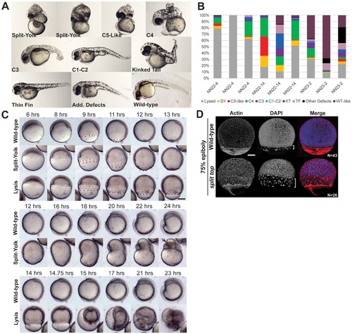
split top mutant embryo phenotypes. (A) 1dpf split top mutant embryo C1 to C4 dorsalized phenotypes, split-yolk (SY), kinked tail (KT) and thin-fin (TF) phenotypes or additional (Add.) defects. (B) Three mutant females (NN22-4, NN22-14 and NN23-2) illustrate that clutches from a single mutant mother exhibit similar phenotypic trends, but also variability in phenotypic distribution. (C) Time-lapse imaging of wild-type and split top mutant embryos was performed at 21°C, thus development proceeded more slowly than at 28°C. Red arrowheads mark the deep cells and black ones, the EVL. Yellow asterisks on the split-yolk mutant mark the yolk and the arrow marks the developing eye. (D) Confocal z-projections of double-stained embryos. In most split top embryos EVL migration is uncoupled from deep cells, as indicated with brackets. Scale bars: 240µm (C), 150µm (D). |
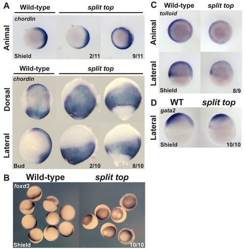
Dorsoventral marker analysis. (A) chordin, (B) foxd3, (C) tolloid and (D) gata2 expression. Animal and lateral views, dorsal to right. Dorsal views, anterior to top. |
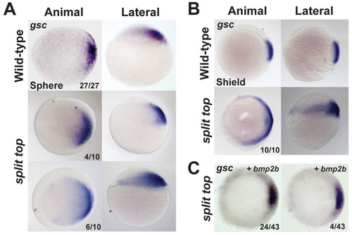
Expanded goosecoid expression in split top mutant embryos. (A) Sphere and (B,C) shield stage embryos. (C) Injection of bmp2b mRNA rescued the expanded gsc expression domain of mutants (animal views). In addition to the embryos with the stainings shown, 15 of 43 embryos showed weak or no gsc expression, indicating ventralization. Dorsal to right. |
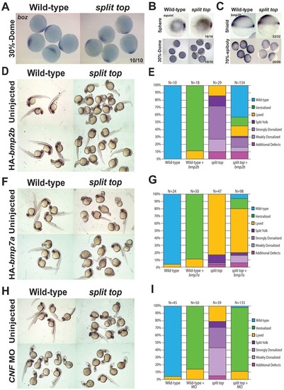
BMP misexpression rescues split top mutants and endogenous BMP signaling is functional. (A) bozozok and (B) squint expression (sphere stage: animal view, dorsal to right). (C) bmp2b expression at shield (lateral view, dorsal to right) and mid-gastrulation. (D-G) Representative phenotypes and bar graphs of uninjected and bmp2b (D,E) or bmp7a (F,G) mRNA injected wild-type and split top mutant embryos. (H,I) Representative phenotypes and bar graphs of wild-type and split top mutant embryos uninjected or injected with chordin, noggin and fstl1b morpholinos (CNF MO). EXPRESSION / LABELING:
|
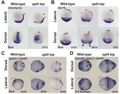
Convergent extension in split top embryos. Expression of (A) brachyury, (B) tbx16, (C,D) pax2.1, (D) krox20 and (C,D) myod indicates an expansion of the midline (probably combined with increased midline mesoderm tissue) and reduced extension in split top mutants. Additionally, whereas krox20 marks rhombomeres 3 and 5 in wild-type embryos, in split top mutants a single krox20 stripe is evident, indicating a loss of either rhombomeres 3 or 5, or a delay in rhombomere 5 expression. Black arrows mark the anterior-most wild-type expression domain and red arrows the anterior-most split top expression domain. The dorsal midline tissue is marked by T-bars in wild-type (black or white) and split top (red) mutants. The brackets indicate the distance from the anterior-most point of the embryo to the neural markers. In A,B, anterior is to top; in C,D, anterior is to left. EXPRESSION / LABELING:
PHENOTYPE:
|
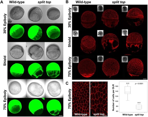
Microtubules and actin cytoskeleton are disrupted. (A) Wild-type and split top mutants embryos immunostained to label microtubules in the YCL (N=4-5 and N=5 embryos at each stage, respectively). (B) Wild-type and split top mutant embryos stained with Phalloidin conjugated to Alexa Fluor 568 marks actin in the YCL (N=4-5 embryos at each stage). Bright field (BF) images. (C) High-magnification confocal z-projections of Phalloidin-stained embryos and box plot showing the wild-type and mutant EVL cell morphology and number, respectively. P value is shown. Scale bars: 280µm (A), 240µm (B), 70µm (C). EXPRESSION / LABELING:
PHENOTYPE:
|
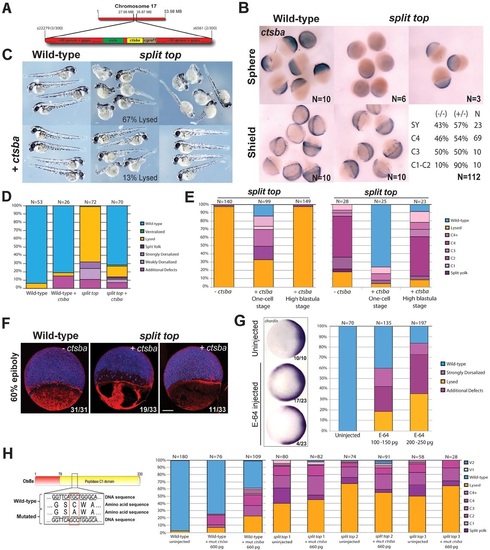
Cathepsin Ba is deficient in split top mutant embryos. (A) Schematic of the split top interval on chromosome 17. (B) ctsba mRNA expression in wild-type and split top mutant embryos. The genotype and strength of the split top mutant phenotype. (C) Representative phenotypes of uninjected and ctsba-injected wild-type and split top mutant embryos, and (D) bar graphs showing rescue. (E) ctsba mRNA injection in the yolk cell of mid-blastula (high to dome stage) embryos from two homozygous split top mutant females did not rescue the lysis or dorsalized phenotypes. (F) Whole-mount 60% epiboly (early gastrula) embryos stained with actin (red) and DAPI (blue). All uninjected split top mutants exhibited large patches of actin-deficient YCL regions. In the right embryo injected with ctsba mRNA, the gap between the YSN/EVL and the deep cells is rescued, along with partial rescue of the actin cytoskeleton; 3 of 33 embryos showed no actin staining. Scale bar: 140µm. (G) E-64-injected wild-type embryos show expanded chordin expression (animal pole view, dorsal to right). Bar graphs show 1dpf phenotypic distributions. (H) Mis-sense mutation made in the catalytic domain of Ctsba. Bar graphs show 1dpf phenotypic distributions of this mutant mRNA injected into wild-type embryos or embryos from three different split top mutant females. |