
- Title
-
Characterization of two novel small molecules targeting melanocyte development in zebrafish embryogenesis
- Authors
- Chen, L., Ren, X., Liang, F., Li, S., Zhong, H., and Lin, S.
- Source
- Full text @ Pigment Cell Melanoma Res.
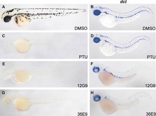
The two compounds 12G9 and 36E9 inhibit pigmentation. The development of 12G9 and 36E9 treated embryos was slightly delayed. To analyze treated embryos at the same stage as control groups, these embryos were collected at 54 hpf. Lateral view, anterior to the left and dorsal to the top. A, C, E, and G show the live embryos; B, D, F, and H show in situ hybridization with dct probe. (A and B) DMSO control group. Treated embryos had stellate melanocytes with corresponding dct expression. (C and D) Embryos were treated with PTU. PTU blocked melanin synthesis and caused unpigmented embryos. However, the expression of dct showed that melanocytes were still present. (E and F) Embryos treated with 12G9. (G and H) Embryos treated with 36E9. Both 12G9 and 36E9 prevented pigmentation, and the number of dct positive cells in the trunk and RPE are much less than that of control group. |

12G9 and 36E9 induced morphological changes in differentiated melanocytes. All embryos were treated from 30 to 48 hpf except in A and B. Lateral view, anterior to the left and dorsal to the top. A, C, E, G, and I show the pictures of live embryos; B, D, F, H, and J show the dct in situ hybridization. (A and B) 30 hpf embryos before treatment. (C and D) Embryos treated with DMSO had stellate melanocytes, and the pattern of dct positive cells matched the melanocytes. (E and F) Embryos treated with PTU had less melanin, and the dct expression pattern was normal. Embryos treated with compound 12G9 (G and H) or 36E9 (I and J) had less pigmentation, condensed melanocytes, and severely reduced number of dct positive cells. |
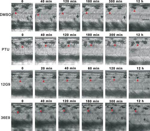
Time-lapse microscopy imaging revealed melanocytes shrank to condensed dots when embryos were treated with 12G9 or 36E9 from 36 to 48 hpf. DMSO and PTU were used as control. All panels show dorsal stripes of trunk region. Time points are indicated on top of each panel. Red arrowheads point to the melanocytes under transformation. |
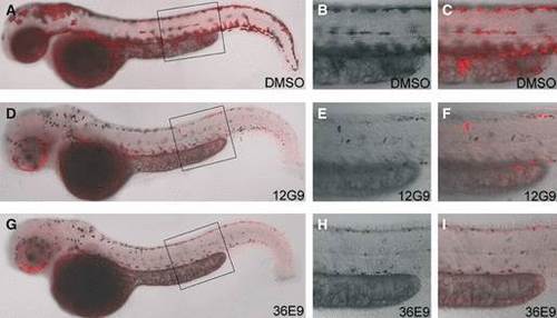
12G9 and 36E9 treatment leads to loss of dct expression in melanocytes. All embryos were treated from 30 to 48 hpf. Lateral view, anterior to the left and dorsal to the top. Dct expression is in red. The right two columns are higher magnifications of boxed areas in the most left column. (A to C) In DMSO treated embryos, melanin (black) and dct expression (red) overlapped perfectly. (D to I) In 12G9 or 36E9 treated embryos, only a few melanocytes had overlapping melanin and dct expression. |
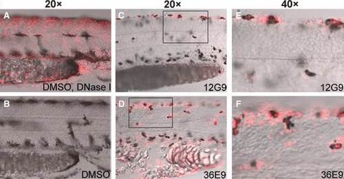
12G9 and 36E9 caused apoptosis of melanocytes. All embryos were treated from 30 to 48 hpf. Lateral view, anterior to the left and dorsal to the top. Red signals show the apoptotic cells detected by TUNEL assay. (A) Positive TUNEL assay control group, fixed embryos were digested with DNase I and then subjected to TUNEL assay. Many cells were stained with red signal in nuclei. (B) DMSO control group, no red TUNEL signals were detected. Embryos treated with 12G9 (C) and 36E9 (D) had overlap of black and red signals, indicating some melanocytes were undergoing apoptosis. (E) and (F) show higher magnification of boxed area in (C) and (D). |
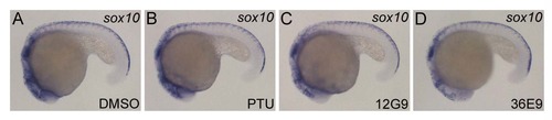
12G9 and 36E9 did not impair neural crest cell development. |
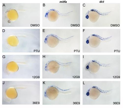
The effects of 12G9 and 36E9 on the development of melanocyte lineage. |
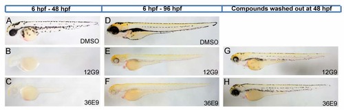
Recovery of melanocyte after removal of 12G9 and 36E9 at 48 hpf. |