
FIGURE
Fig. 4
- ID
- ZDB-FIG-151112-9
- Publication
- Chen et al., 2012 - Characterization of two novel small molecules targeting melanocyte development in zebrafish embryogenesis
- Other Figures
- All Figure Page
- Back to All Figure Page
Fig. 4
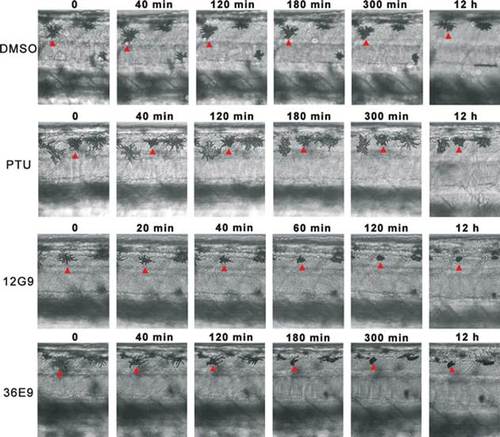
Time-lapse microscopy imaging revealed melanocytes shrank to condensed dots when embryos were treated with 12G9 or 36E9 from 36 to 48 hpf. DMSO and PTU were used as control. All panels show dorsal stripes of trunk region. Time points are indicated on top of each panel. Red arrowheads point to the melanocytes under transformation. |
Expression Data
Expression Detail
Antibody Labeling
Phenotype Data
Phenotype Detail
Acknowledgments
This image is the copyrighted work of the attributed author or publisher, and
ZFIN has permission only to display this image to its users.
Additional permissions should be obtained from the applicable author or publisher of the image.
Full text @ Pigment Cell Melanoma Res.