
|
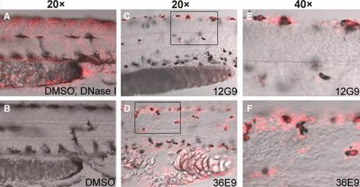
Fig. 6
12G9 and 36E9 caused apoptosis of melanocytes. All embryos were treated from 30 to 48 hpf. Lateral view, anterior to the left and dorsal to the top. Red signals show the apoptotic cells detected by TUNEL assay. (A) Positive TUNEL assay control group, fixed embryos were digested with DNase I and then subjected to TUNEL assay. Many cells were stained with red signal in nuclei. (B) DMSO control group, no red TUNEL signals were detected. Embryos treated with 12G9 (C) and 36E9 (D) had overlap of black and red signals, indicating some melanocytes were undergoing apoptosis. (E) and (F) show higher magnification of boxed area in (C) and (D).