
- Title
-
Recapitulation of zebrafish sncga expression pattern and labeling the habenular complex in transgenic zebrafish using green fluorescent protein reporter gene
- Authors
- Chen, Y.C., Cheng, C.H., Chen, G.D., Hung, C.C., Yang, C.H., Hwang, S.P., Kawakami, K., Wu, B.K., and Huang, C.J.
- Source
- Full text @ Dev. Dyn.
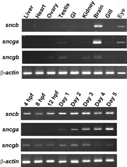
Expression profiles of zebrafish synuclein-related transcripts in various adult tissues and at different developmental stages by reverse transcriptase-polymerase chain reaction (RT-PCR). A,B: RT-PCR was performed with gene-specific primers. β-actin bands were used to normalize the amount of cDNA prepared from different tissues (A) and from different developmental stages (B). The developmental expression profile of each gene was examined in embryos from 4 hours postfertilization (hpf) to 5 dpf. |
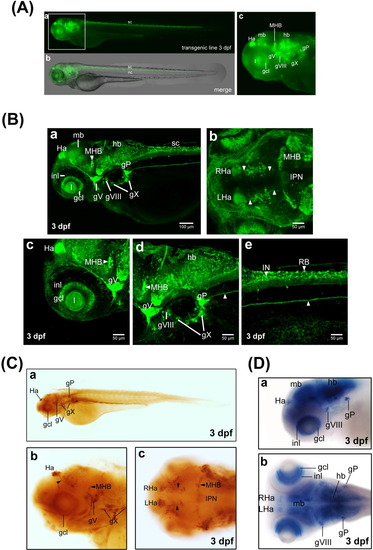
The expression pattern of green fluorescent protein (GFP) in transgenic zebrafish Tg(sncga:GFP) line. Microinjection of the expression construct into zebrafish embryos at the one- to two-cell stage and generation of transgenic GFP line by means of Tol2-mediated transgenesis were described in the text. A: Images from Tg(sncga:GFP) transgenic line at 3 days postfertilization (dpf). Images of bright field and fluorescence are merged and shown in panel b, while fluorescence images are shown in panel a. sc, spinal cord; nc, notochord. B: Confocal image analysis. Zebrafish larva of transgenic Tg(sncga:GFP) line at 3 dpf were collected and fixed with 4% paraformaldehyde. After treatment of FocusClear, embryos were subjected to high-resolution confocal image analysis. All embryos are shown with the anterior to the left. Embryos in a, c, d, e are shown in lateral view, while embryos in b are shown in dorsal view. White arrowheads indicated habenulo-interpeduncular projection (panel b) and posterior lateral line projection (panels d, e). C: Whole-mount immunostaining of Tg(sncga:GFP) embryos by polyclonal antibody against GFP. Embryos in a and b are shown in lateral view, while embryos in c are shown in dorsal view. The habenulo-interpeduncular projection was labeled by black arrowheads. D: Expression patterns of sncga mRNA in zebrafish embryo at 3 dpf. gcl, retinal ganglion cell layer; gP, posterior lateral line ganglion; gV, trigeminal ganglion; gVIII, statoacoustic ganglion; gX, vagal ganglion; Ha, habenula; hb, hindbrain; mb, midbrain; IN, interneuron; inl, inner nuclear layer; IPN, interpeduncular nucleus; l, lens; LHa, left-side habenula; MHB, midbrain-hindbrain boundary; RB, Rohon-Beard neuron; RHa, right-side habenula. EXPRESSION / LABELING:
|
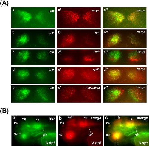
Double fluorescence in situ hybridizations of habenula-expressing markers, gfp and sncga in Tg(sncga:GFP) fish. A: Expression domains of sncga (a′), lov (b′), ron (c′), cpd2 (d′) and f-spondin2 (e′) along with gfp (a-e) in habenula in Tg(sncga:GFP) fish at 3 days postfertilization (dpf). a″-e″, merged image of a and a′, b and b′, c and c′; d and d′, and e and e′, respectively. B: Double fluorescence in situ hybridization of sncga and gfp in an Tg(sncga:GFP) fish at 3 dpf. Panel a shows the staining of gfp mRNA, whereas panel b shows the staining of sncga mRNA. Panel c shows the merged image, in which gfp and sncga staining completely overlapped. gcl, retinal ganglion cell layer; GFP, green fluorescent protein; gP, posterior lateral line ganglion; Ha, habenula; hb, hindbrain; l, lens; mb, midbrain. EXPRESSION / LABELING:
|