- Title
-
Robo2 is required for establishment of a precise glomerular map in the zebrafish olfactory system
- Authors
- Miyasaka, N., Sato, Y., Yeo, S.Y., Hutson, L.D., Chien, C.B., Okamoto, H., and Yoshihara, Y.
- Source
- Full text @ Development
|
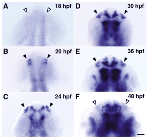
Transient expression of robo2 mRNA in the developing olfactory placode, shown by whole-mount in situ hybridization. (A-E) Dorsal views with anterior to the top; (F) ventral view with anterior to the top. As the olfactory placode moves from a dorsal to a more ventral location during development, it is more easily viewed from the ventral side at 48 hpf. Arrowheads (both open and closed) denote the position of the olfactory placode. robo2 mRNA was detected in the olfactory placode in embryos at stages between 20-36 hpf (closed arrowheads in B-E). Scale bar: 50 µm. EXPRESSION / LABELING:
|
|
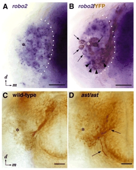
The unipolar neurons express robo2 and make pathfinding errors in ast embryos. (A,B) Frontal views of the olfactory placode of 30-hpf omp:yfp/+ embryos. (A) Whole-mount in situ hybridization. Blue dots are signals for robo2 mRNA on the focal plane. Broad blue staining in the olfactory placode is derived from signals on cells that are out of the focal plane. (B) Double stainning for robo2 mRNA and YFP antigen. Early developing OSNs (arrows) and unipolar neurons (arrowheads) are stained in brown with anti-GFP antibody. Hybridization signals for robo2 mRNA (blue dots) are seen on the somata of both cell types. White dotted lines indicate the boundary between the olfactory placode and telencephalon. (C,D) Frontal views of the head of wild-type (C) and ast homozygous (D) embryos stained with zns-2 antibody at 36 hpf. The zns-2-positive axons misroute medially or ventromedially immediately after exiting the olfactory placode in ast embryos (arrows in D). Asterisks mark the position of the olfactory pit. d, dorsal; m, medial. Scale bar: 50 µm. EXPRESSION / LABELING:
|
|
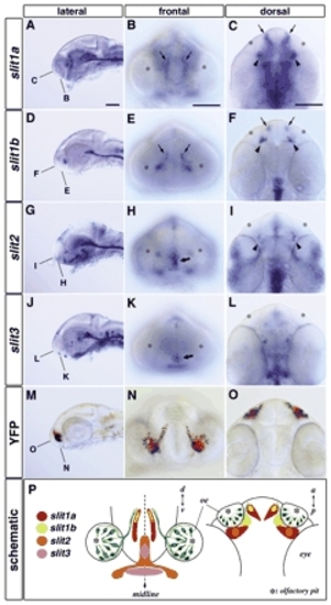
Spatial expression patterns of four Slit mRNAs are consistent with a function as repulsive cues for the olfactory axons. Heads of 30-hpf whole-mounted embryos hybridized with slit1a (A-C), slit1b (D-F), slit2 (G-I) and slit3 (J-L) probes. The olfactory axon trajectory of a Tg(OMP2k:gap-YFP)rw032a transgenic embryo stained with anti-GFP antibody is shown in M-O. slit1a, slit1b and slit2 are expressed in bilateral clusters of cells located near the boundary between the telencephalon and diencephalon (arrowheads in C,F,I). slit1a and slit1b are also expressed bilaterally in the telencephalon (arrows in B,C,E,F). slit2 and slit3 are expressed along the midline in the ventral forebrain (thick arrows in H,K). The correlation between the regions of Slit expression and the olfactory axon trajectory (green) is schematized in P. A,D,G,J,M, lateral views with anterior to the left; B,E,H,K,N, frontal views with dorsal to the top; C,F,I,L,O, dorsal views with anterior to the top. The focal planes of frontal (B,E,H,K,N) and dorsal (C,F,I,L,O) views are indicated in the corresponding leftmost panels (A,D,G,J,M). Asterisks mark the position of the olfactory pit. Scale bar: 100 µm. |
|
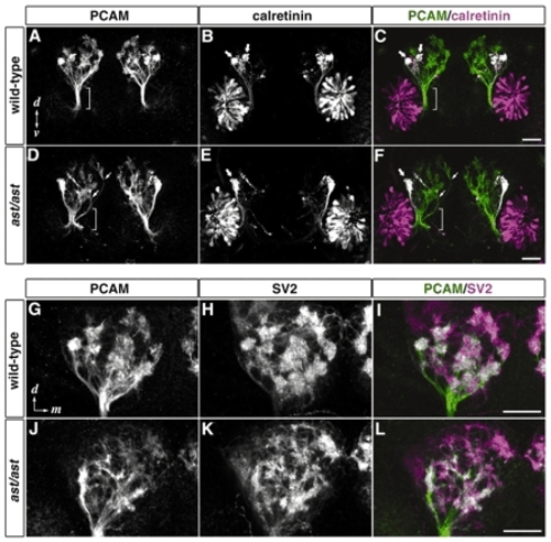
ast embryos show defasciculation of the olfactory nerve and impaired proto-glomerular organization. Frontal views of the head of wild-type (A-C,G-I) and ast homozygous (D-F,J-L) 72-hpf embryos stained with antibodies against PCAM (A,D,G,J; green in C,F,I,L), calretinin (B,E; magenta in C,F) and SV2 (H,K; magenta in I,L). (A-F) In wild type, olfactory axons maintain a tightly fasciculated state until they enter the OB (brackets in A,C). By contrast, olfactory axons in ast are defasciculated before reaching the OB (brackets in D,F) and some fibers enter the OB from improper entry sites (arrows in D,F). Calretinin-positive axons mainly project laterally to form two discrete proto-glomeruli in wild type (thick arrows in B,C), whereas in ast, only one irregularly shaped proto-glomerulus is seen at the lateralmost position in the OB (thick arrows in E,F). (G-L) Proto-glomeruli stained with antibodies against PCAM and SV2 in ast embryos (J-L) are less clearly defined than those in wild type (G-I). d, dorsal; v, ventral; m, medial. Scale bar: 50 µm.
EXPRESSION / LABELING:
|

Unillustrated author statements |


