
Fig. 7
- ID
- ZDB-FIG-050513-14
- Publication
- Miyasaka et al., 2005 - Robo2 is required for establishment of a precise glomerular map in the zebrafish olfactory system
- Other Figures
- All Figure Page
- Back to All Figure Page
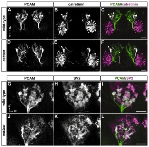
ast embryos show defasciculation of the olfactory nerve and impaired proto-glomerular organization. Frontal views of the head of wild-type (A-C,G-I) and ast homozygous (D-F,J-L) 72-hpf embryos stained with antibodies against PCAM (A,D,G,J; green in C,F,I,L), calretinin (B,E; magenta in C,F) and SV2 (H,K; magenta in I,L). (A-F) In wild type, olfactory axons maintain a tightly fasciculated state until they enter the OB (brackets in A,C). By contrast, olfactory axons in ast are defasciculated before reaching the OB (brackets in D,F) and some fibers enter the OB from improper entry sites (arrows in D,F). Calretinin-positive axons mainly project laterally to form two discrete proto-glomeruli in wild type (thick arrows in B,C), whereas in ast, only one irregularly shaped proto-glomerulus is seen at the lateralmost position in the OB (thick arrows in E,F). (G-L) Proto-glomeruli stained with antibodies against PCAM and SV2 in ast embryos (J-L) are less clearly defined than those in wild type (G-I). d, dorsal; v, ventral; m, medial. Scale bar: 50 µm.
|
| Gene: | |
|---|---|
| Fish: | |
| Anatomical Term: | |
| Stage: | Protruding-mouth |