
- Title
-
Spatial and temporal regulation of ventral spinal cord precursor specification by Hedgehog signaling
- Authors
- Park, H.C., Shin, J., and Appel, B.
- Source
- Full text @ Development
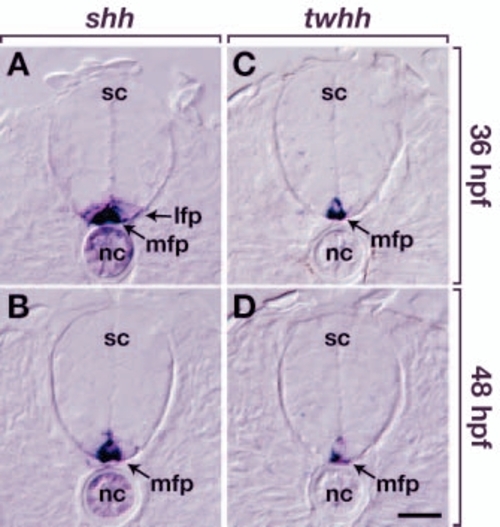
Zebrafish embryos express Hh ligands during the period of oligodendrocyte specification. All panels show transverse sections through trunk spinal cord (sc), with dorsal upwards. (A) Notochord (nc), medial floor plate (mfp) and lateral floor plate (lfp) express shh at 36 hfp. (B) At 48 hpf, medial floor plate and notochord express shh. (C) Medial floor plate also expresses twhh at 36 hpf and (D) 48 hpf. Scale bar: 20 µm. |
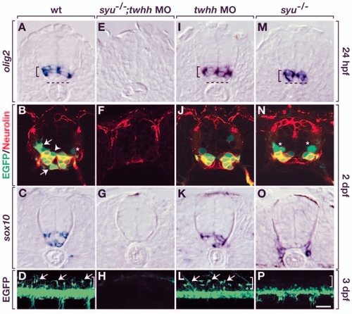
Motoneurons and oligodendrocytes have genetically separable requirements for Hh signaling. Top three rows of panels show transverse sections through trunk spinal cord, with dorsal upwards. Bottom row shows lateral views of intact olig2:egfp embryos, with dorsal upwards and anterior towards the left. (A) Wild-type embryos expressed olig2 RNA in the ventral spinal cord (bracket), but the ventralmost cells were olig2–. Broken line indicates the ventral boundary of the spinal cord. (B) olig2:EGFP+ cells of wild-type embryos included Neurolin+ SMNs, OPCs (arrows), Neurolin– cells near ventricle (arrowhead) and a faint GFP+, Neurolin– cell near pial surface (asterisk), which could be a primary motoneuron or VeLD interneuron. (C) sox10+ OPCs in wild-type embryo. (D) Arrows indicate dorsally migrated OPCs in wild-type embryo. Bracket indicates dorsal spinal cord. (E-H) syu–/– embryos injected with twhh MO lacked olig2+ cells (E), secondary motoneurons (F) and OPCs (G,H). (I-L) Wild-type embryos injected with twhh MO expressed olig2 RNA (I) and produced secondary motoneurons (J) and OPCs (K,L). syu–/– embryos expressed olig2 RNA (M), but olig2+ cells were located more ventrally than normal. (M-P) syu–/– embryos had olig2:EGFP+, Neurolin+ secondary motoneurons and olig2:EGFP+, Neurolin– cells (asterisks) (N); however, they did not have OPCs (O,P). (O) sox10+ cells are probably Schwann cells (Dutton et al., 2001), which do not appear in all sections. Scale bar: 20 µm for upper three rows; 40 µm for bottom row. EXPRESSION / LABELING:
|
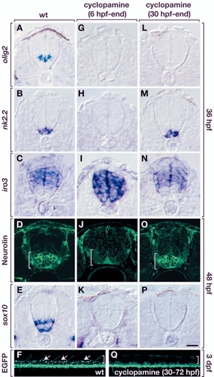
Hh signaling is required for oligodendrocyte specification after dorsoventral spinal cord patterning and motoneuron development. All panels in the top five rows show transverse sections, dorsal upwards, of trunk spinal cord. Bottom two panels are side views of whole embryos, dorsal upwards and anterior leftwards. (A-E) Control embryos showing normal expression of various markers. (G-K) Embryos treated with cyclopamine from 6 hpf onwards did not express olig2 (G) or nkx2.2 (H), and expressed iro3 in ventral spinal cord (I), indicating that ventral spinal cord patterning was lost. These embryos did not produce SMNs (J) or OPCs (K). (L-Q) Embryos treated with cyclopamine from 30 hpf onward did not express olig2 by 36 hpf (L) but expressed nkx2.2 (M) and iro3 (N) in their normal patterns. SMNs were produced in normal numbers (O) but OPCs were absent (P,Q). (F) Untreated Tg[olig2:egfp] embryo showing OPCs (arrows) in dorsal spinal cord (brackets). Scale bar: 20 µm for top five rows; 80 µm for F and Q. EXPRESSION / LABELING:
|
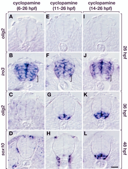
Conditional manipulation of Hh signaling reveals a crucial period for specification of oligodendrocyte precursors. All panels show transverse sections, dorsal side upwards. (A-D) Embryos incubated in cyclopamine from 6-26 hpf. At 26 hpf, these embryos did not express olig2 (A). iro3 expression included the ventralmost spinal cord cells (B; brackets in B,F,J). olig2 expression was not recovered by 36 hpf (C) and no sox10+ OPCs were evident by 48 hpf (D). (E-H) Embryos treated with cyclopamine from 11-26 hpf. olig2 expression was absent by 26 hpf (E) but a small domain of iro3– cells was present in the ventral spinal cord (F). These embryos recovered olig2 expression by 36 hpf, but in an abnormally ventral position (G). At 48 hpf, few sox10+ OPCs had developed (H). (I-L) Embryos treated with cyclopamine from 14-26 hpf. olig2 expression was not maintained until 26 hpf (I) but iro3 expression appeared normal (J, compare with Fig. 6C). olig2 expression was recovered by 36 hpf in ventralmost spinal cord cells (K) but in a larger domain compared with 11-26 hpf treated embryos. These embryos expressed sox10 at 48 hpf, but in an abnormally ventral position (L, compare with Fig. 6E). Scale bar: 20 µm. |