
|
OPTICS: MAGNIFICATION: DATE OF IMAGE: SUBMITTER COMMENTS:
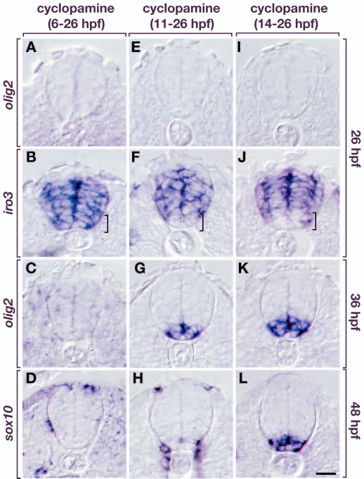
Fig. 7 Conditional manipulation of Hh signaling reveals a crucial period for specification of oligodendrocyte precursors. All panels show transverse sections, dorsal side upwards. (A-D) Embryos incubated in cyclopamine from 6-26 hpf. At 26 hpf, these embryos did not express olig2 (A). iro3 expression included the ventralmost spinal cord cells (B; brackets in B,F,J). olig2 expression was not recovered by 36 hpf (C) and no sox10+ OPCs were evident by 48 hpf (D). (E-H) Embryos treated with cyclopamine from 11-26 hpf. olig2 expression was absent by 26 hpf (E) but a small domain of iro3– cells was present in the ventral spinal cord (F). These embryos recovered olig2 expression by 36 hpf, but in an abnormally ventral position (G). At 48 hpf, few sox10+ OPCs had developed (H). (I-L) Embryos treated with cyclopamine from 14-26 hpf. olig2 expression was not maintained until 26 hpf (I) but iro3 expression appeared normal (J, compare with Fig. 6C). olig2 expression was recovered by 36 hpf in ventralmost spinal cord cells (K) but in a larger domain compared with 11-26 hpf treated embryos. These embryos expressed sox10 at 48 hpf, but in an abnormally ventral position (L, compare with Fig. 6E). Scale bar: 20 µm.
| Preparation | Image Form | View | Direction |
| not specified | still | not specified | not specified |