
Fig. 6
|
OPTICS: MAGNIFICATION: DATE OF IMAGE: SUBMITTER COMMENTS:
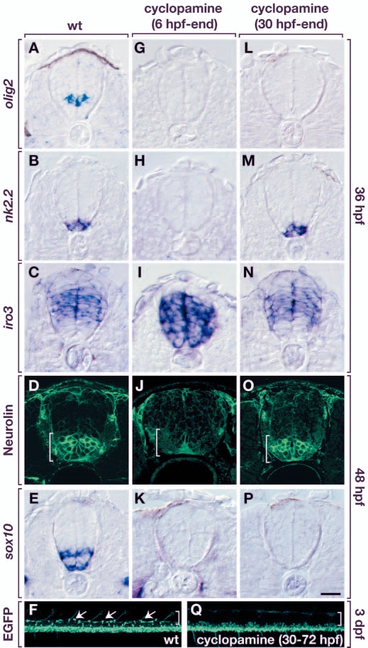
Fig. 6 Hh signaling is required for oligodendrocyte specification after dorsoventral spinal cord patterning and motoneuron development. All panels in the top five rows show transverse sections, dorsal upwards, of trunk spinal cord. Bottom two panels are side views of whole embryos, dorsal upwards and anterior leftwards. (A-E) Control embryos showing normal expression of various markers. (G-K) Embryos treated with cyclopamine from 6 hpf onwards did not express olig2 (G) or nkx2.2 (H), and expressed iro3 in ventral spinal cord (I), indicating that ventral spinal cord patterning was lost. These embryos did not produce SMNs (J) or OPCs (K). (L-Q) Embryos treated with cyclopamine from 30 hpf onward did not express olig2 by 36 hpf (L) but expressed nkx2.2 (M) and iro3 (N) in their normal patterns. SMNs were produced in normal numbers (O) but OPCs were absent (P,Q). (F) Untreated Tg[olig2:egfp] embryo showing OPCs (arrows) in dorsal spinal cord (brackets). Scale bar: 20 µm for top five rows; 80 µm for F and Q.
| Preparation | Image Form | View | Direction |
| not specified | still | not specified | not specified |