
Fig. 5
- ID
- ZDB-FIG-250813-26
- Publication
- Halford et al., 2025 - TMEM63A, associated with hypomyelinating leukodystrophies, is an evolutionarily conserved regulator of myelination
- Other Figures
- All Figure Page
- Back to All Figure Page
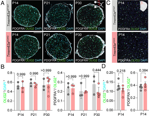
Loss of Tmem63a does not affect OPC and OL populations. (A) Representative micrographs of 14 µm-thick optic nerve sections immunostained against OLIG2 (green) and PDGFRA (gray), and counterstained with DAPI (cyan) from Tmem63aWT/WT and Tmem63aKO/KO mice at P14, P21, and P30. (Scale bar, 100 µm.) (B) Left, percentage OLIG2+ cells/DAPI+ cells in the optic nerve for Tmem63aWT/WT and Tmem63aKO/KO mice. Right, percentage PDGFRA+ cells/OLIG2+ cells in the whole cross section of the optic nerve for Tmem63aWT/WT and Tmem63aKO/KO mice (n = 3 to 5 animals per genotype and age, Brown–Forsythe and Welch ANOVA). (C) Representative micrographs of 16 µm-thick cortical sections immunostained against OLIG2 (green) and PDGFRA (gray), and counterstained with DAPI (cyan) from Tmem63aWT/WT and Tmem63aKO/KO mice at P14. (Scale bar, 100 µm.) (D) Left, percentage OLIG2+ cells/DAPI+ cells in the cortex for Tmem63aWT/WT and Tmem63aKO/KO mice. Right, percentage PDGFRA+ cells/OLIG2+ cells in the cortex for Tmem63aWT/WT and Tmem63aKO/KO mice (n = 4 animals per genotype, unpaired t test). |