|
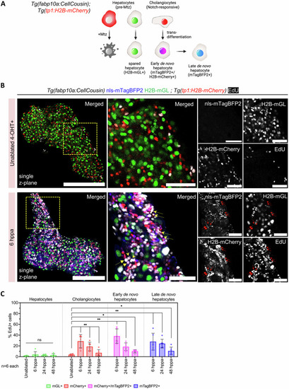
Cholangiocytes proliferate in response to partial ablation of hepatocytes. A Schematic illustrating the lineage tracing of cholangiocytes after partial ablation via the CellCousin method. Hepatocytes are labeled with H2B-mGL+ or mCherry-NTR+ after 4-OHT treatment in the Tg(fabp10a:CellCousin) line. Cholangiocytes, which are Notch-responsive, are marked with bright H2B-mCherry+ (nuclear red) using the Tg(tp1:H2B-mCherry) line. After MTZ administration, Specific ablation of mCherry-NTR+ hepatocytes leads to a liver with spared H2B-mGL+ hepatocytes and de novo nls-mTagBFP2+ hepatocytes. Early de novo hepatocytes derived from cholangiocytes are detected as nls-mTagBFP2 + /H2B-mCherry+ (nuclear magenta), while late de novo hepatocytes, due to dilution of H2B-mCherry label, are marked only with nls-mTagBFP2. B Confocal images of livers from EdU labeled uninjured and 6 hppa Tg(fabp10a:CellCousin); Tg(tp1:H2B-mCherry) transgenic animals. Arrows mark proliferating early de novo hepatocytes. Scale bar: 200 μm (left panels), 50 μm (insets). C Barplot with Mean ± SD showing quantification of the percentage of EdU+ hepatocytes, cholangiocytes, and early and late de novo hepatocytes in uninjured, 6 dppa, 24 dppa and 48 dppa (n = 6 animals). (ANOVA, followed by pair wise comparison using Two-Tailed Mann Whitney. For cholangiocytes and de novo hepatocytes, comparison with uninjured cholangiocytes is shown. ** p-value = 0.0022; * p-value < 0.05).
|