
Fig. 3
- ID
- ZDB-FIG-250428-129
- Publication
- Hu et al., 2025 - Dscamb regulates cone mosaic formation in zebrafish via filopodium-mediated homotypic recognition
- Other Figures
- All Figure Page
- Back to All Figure Page
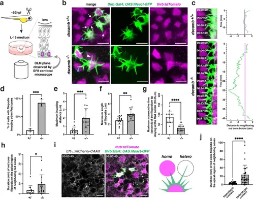
Dscamb is required for filopodium-mediated homotypic recognition.a Experimental design for ex vivo live imaging. OLM: outer limiting membrane. b Confocal images of the apical surface of a cone photoreceptor layer with two transgenes Tg[thrb:Gal4VP16; UAS:lifeact-GFP] and Tg[thrb:tdTomato], which visualize apical filopodia (green) and cell bodies (magenta) of red cones, respectively. In wild type, red cones extend multiple filopodia toward neighboring cones. However, filopodia stop growing when they meet neighboring red cones (white/blue arrows). On the other hand, in dscamb mutants, filopodia keep growing even after they contact other red cones (white/blue arrowheads). A blue arrow and arrowhead indicate wild-type and dscamb mutant filopodia used for time-lapse analysis shown in the panel c. Three independent experiments. c Time-lapse observation of filopodial behavior in wild-type and dscamb mutant red cones. Right histograms show a temporal profile of the distance between a filopodial tip and an apical domain border of neighboring red cones, which was measured every 20.6 s. d Percentage of red cones with invading filopodia. e Maximum invading distance of apical filopodia. f Maximum length of apical filopodia. g Maximum consecutive time of filopodium tip staying within the border zone ( ± 0.25 μm) of the apical domain of red cones. h Residence time of red cone filopodia on apical domains of neighboring red cones. i Confocal scanning of the photoreceptor layer in triple transgenic Tg[thrb:Gal4VP16; UAS:lifeact-GFP; Ef1α:mCherry-CAAX] retinas, which visualize red cone filopodia (green) contacting on other red cones (homotypic, magenta) or other non-red cone-type photoreceptors (heterotypic, white). j Residence time of red cone filopodia (collected from 3 wild-type samples) on apical domains of other red cones (homotypic contact) or other non-red cone-type photoreceptors (heterotypic contact). d–h, j Means ± SD. Statistical significance was evaluated with unpaired t-tests (two tail): *p < 0.0332, **p < 0.0021, ***p < 0.0002, and ****p < 0.0001. Biological replicates: d Three embryos. e–h n = 13 for wild-type filopodia and n = 14 for dscamb mutant filopodia. j n = 30 for filopodia showing homotypic contact and n = 26 for filopodia showing heterotypic contact. Scale bars: 5 μm b, i. d–h, j Source data and p-values are provided as Source Data for Fig. 3. |