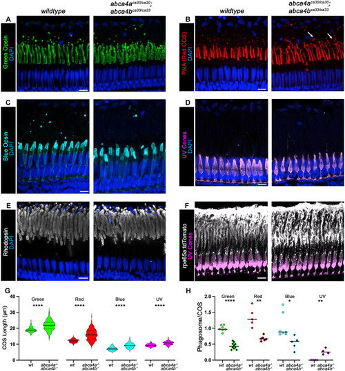
Photoreceptors, RPE and phagosomes in retina of young juvenile wild-type or abca4 double mutant zebrafish. (A-F) High-resolution confocal z-projections of retina obtained from wild-type or double abca4aca30/ca30;abca4bca33/ca33 mutant zebrafish at 5 weeks of age. Images show (A) green cone outer segments (COS) stained for Green Opsin (green, z=3.24 µm); (B) red COS labeled with PNA (PNA, z=4.5 µm) with distal tip bulges indicated by white arrows; (C) blue COS stained for Blue Opsin (cyan, z=4.5 µm); (D) UV cones expressing EGFP (violet, z=5 µm); (E) rod outer segments (ROS) stained for rhodopsin (white, z=2.7 µm) and (F) retinal pigmented epithelium (RPE) expressing tdTomato labeled for RFP (white, z=3.24 µm). Nuclei are shown in blue (DAPI). Scale bars: 10 µm. (G) Quantification of COS length. Solid horizontal lines represent the mean and dashed horizontal lines indicate quartiles. (H) Quantification of shed phagosomes per COS. Solid horizontal line represents the mean. *P<0.05; **P<0.01; ****P<0.0001 (Welch's t-test).
|
