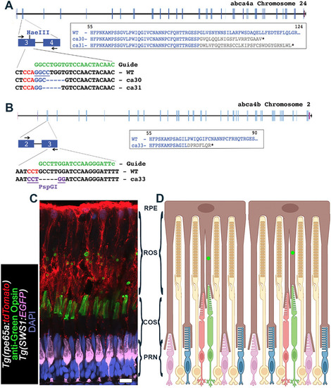
Generation of zebrafish abca4 mutants by using CRISPR/Cas9. (A) Shown is a sequence in exon 3 of the abca4a gene on chromosome 24 (NCBI: Gene ID: 798993) that was targeted with a synthetic guide RNA. Two mutant alleles – abca4aca30 (ca30) and abca4aca31 (ca31) − that disrupt a HaeIII restriction site, were identified and mutant lines established. Both mutant alleles disrupt the reading frame through addition of 21 (ca30) or 31 (ca31) missense residues, thereby truncating at a position equivalent to amino acid (aa) 112 or 122 in WT. (B) Shown is a sequence in exon 3 of the abca4b gene on chromosome 2 (NCBI: Gene ID: 555506) that was targeted with a synthetic guide RNA. The single mutant allele abca4bca33 (ca33) that creates a novel PspGI restriction site, was identified and the mutant line established. Allele ca33 is predicted to disrupt the reading frame through addition of eight missense aa, before truncating at a position equivalent to aa 75 in WT. Synthetic guide RNA sequences are shown in green; Cas9 protospacer adjacent motif (PAM) sequences are shown in red. (C) Confocal z-projection (z=2.82 µm) of the photoreceptor layer in the retina of Tg(rpe65a:tdTomato);Tg(SWS1:EGFP) zebrafish aged 1 year. The retinal pigmented epithelium (RPE) expressing tdTomato (red), UV cones express EGFP (pseudo-colored violet). Green cone outer segment (COS) regions were stained for Green Opsin (green); phagosomes of green COS ingested by RPE microvilli are visible as green ‘spheres’ distal to green COS, which are transported to the RPE cell bodies for digestion. Nuclei are shown in blue (DAPI). Scale bar: 10 µm. (D) Schematic of the photoreceptor layer showing photoreceptor types (yellow rods, green cones, red cones, blue cones, UV cones), RPE (brown), rod outer segment (ROS) region, cone outer segment (COS) region and photoreceptor nuclei (PRN), i.e. outer nuclear region (ONL) region in zebrafish retina. Green dots indicate phagosomes of green COS ingested by RPE microvilli.
|
