|
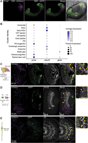
Immunofluorescence identifies enhancer-active cell types. (A) Coronal-orientation image of a NRE-eGFP/HS5-mCherry embryo at 48 hpf showing the activity of NRE (eGFP) in the distal CMZ (stem cell niche) and cells of the INL, and HS5 (mCherry) activity in the temporal retina, in the proximal CMZ, and cells of the INL. (B) Dot plot showing average expression and percentage of cells expressing pcna, elavl3 (encoding HuC/D), and glula (encoding glutamine synthetase [GS]) in cell type clusters. (C) Immunofluorescence for PCNA, mCherry, and eGFP on a coronal eye section from an NRE-eGFP/HS5-mCherry F1 embryo at 48 hpf. PCNA is a marker for progenitors and stem cells in the CMZ. An arrow indicates an mCherry/PCNA-positive cell. An arrowhead indicates a eGFP/PCNA-positive cell. (D) As in (C), but using an antibody detecting HuC/D (elavl3/4). HuC/D is a marker for RGCs in the GCL and amacrine cells in the INL. Arrowheads indicate eGFP/HuC/D-positive cells. (E) As in (C) but using an antibody detecting GS, on an embryo at 72 hpf (sagittal section). GS is a marker for Müller glia. Arrowheads indicate mCherry/GS-positive cells. Scale bars 50 μm, 20 μm in zoom.
|